6EI6 | pdb_00006ei6

CC2D1B coordinates ESRCT-III activity during the mitotic reformation of the nuclear envelope


Experimental Data Snapshot

  • Method: X-RAY DIFFRACTION
  • Resolution: 2.46 Å
  • R-Value Free: 
    0.247 (Depositor), 0.247 (DCC) 
  • R-Value Work: 
    0.205 (Depositor), 0.207 (DCC) 
  • R-Value Observed: 
    0.207 (Depositor) 

wwPDB Validation 3D Report Full Report

Validation slider image for 6EI6

This is version 1.2 of the entry. See complete history

Literature

CC2D1B Coordinates ESCRT-III Activity during the Mitotic Reformation of the Nuclear Envelope.

Ventimiglia, L.N.Cuesta-Geijo, M.A.Martinelli, N.Caballe, A.Macheboeuf, P.Miguet, N.Parnham, I.M.Olmos, Y.Carlton, J.G.Weissenhorn, W.Martin-Serrano, J.

(2018) Dev Cell 47: 547-563.e6

  • DOI: https://doi.org/10.1016/j.devcel.2018.11.012
  • Primary Citation Related Structures: 
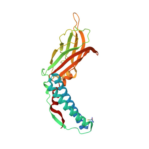
    6EI6

  • PubMed Abstract: 

    The coordinated reformation of the nuclear envelope (NE) after mitosis re-establishes the structural integrity and the functionality of the nuclear compartment. The endosomal sorting complex required for transport (ESCRT) machinery, a membrane remodeling pathway that is highly conserved in eukaryotes, has been recently involved in NE resealing by mediating the annular fusion of the nuclear membrane (NM). We show here that CC2D1B, a regulator of ESCRT polymerization, is required to re-establish the nuclear compartmentalization by coordinating endoplasmic reticulum (ER) membrane deposition around chromatin disks with ESCRT-III recruitment to the reforming NE. Accordingly, CC2D1B determines the spatiotemporal distribution of the CHMP7-ESCRT-III axis during NE reformation. Crucially, in CC2D1B-depleted cells, ESCRT activity is uncoupled from Spastin-mediated severing of spindle microtubules, resulting in persisting microtubules that compromise nuclear morphology. Therefore, we reveal CC2D1B as an essential regulatory factor that licenses the formation of ESCRT-III polymers to ensure the orderly reformation of the NE.


  • Organizational Affiliation
    • Department of Infectious Diseases, King's College London, Faculty of Life Sciences & Medicine, London SE1 9RT, UK.

Macromolecule Content 

  • Total Structure Weight: 63.26 kDa 
  • Atom Count: 4,169 
  • Modeled Residue Count: 484 
  • Deposited Residue Count: 540 
  • Unique protein chains: 1

Macromolecules

Find similar proteins by:|  3D Structure
Entity ID: 1
MoleculeChains  Sequence LengthOrganismDetailsImage
Coiled-coil and C2 domain-containing protein 1-like
A, B
270Drosophila melanogasterMutation(s): 0 
Gene Names: l(2)gd1lgdCG4713
Membrane Entity: Yes 
UniProt
Find proteins for Q9VKJ9 (Drosophila melanogaster)
Explore Q9VKJ9 
Go to UniProtKB:  Q9VKJ9
Entity Groups
Sequence Clusters30% Identity50% Identity70% Identity90% Identity95% Identity100% Identity
UniProt GroupQ9VKJ9
Sequence Annotations
Expand
Reference Sequence

Small Molecules

Ligands 2 Unique
IDChains Name / Formula / InChI Key2D Diagram3D Interactions
PEG

Query on PEG


Download:Ideal Coordinates CCD File
F [auth A]DI(HYDROXYETHYL)ETHER
C4 H10 O3
MTHSVFCYNBDYFN-UHFFFAOYSA-N
SO4

Query on SO4


Download:Ideal Coordinates CCD File
C [auth A]
D [auth A]
E [auth A]
G [auth B]
H [auth B]
C [auth A],
D [auth A],
E [auth A],
G [auth B],
H [auth B],
I [auth B],
J [auth B]
SULFATE ION
O4 S
QAOWNCQODCNURD-UHFFFAOYSA-L

Experimental Data & Validation

Experimental Data

  • Method: X-RAY DIFFRACTION
  • Resolution: 2.46 Å
  • R-Value Free:  0.247 (Depositor), 0.247 (DCC) 
  • R-Value Work:  0.205 (Depositor), 0.207 (DCC) 
  • R-Value Observed: 0.207 (Depositor) 
Space Group: P 1 21 1
Unit Cell:
Length ( Å )Angle ( ˚ )
a = 87.41α = 90
b = 54.05β = 99.52
c = 97.9γ = 90
Software Package:
Software NamePurpose
PHENIXrefinement
MOSFLMdata reduction
SCALAdata scaling
Auto-Rickshawphasing

Structure Validation

View Full Validation Report


Entry History 

Revision History  (Full details and data files)

  • Version 1.0: 2018-10-10
    Type: Initial release
  • Version 1.1: 2018-12-19
    Changes: Data collection, Database references, Structure summary
  • Version 1.2: 2024-10-16
    Changes: Data collection, Database references, Structure summary