6EDR | pdb_00006edr

Crystal Structure of Human CD38 in Complex with 4'-Thioribose NAD+


Experimental Data Snapshot

  • Method: X-RAY DIFFRACTION
  • Resolution: 2.40 Å
  • R-Value Free: 
    0.262 (Depositor), 0.265 (DCC) 
  • R-Value Work: 
    0.216 (Depositor), 0.220 (DCC) 
  • R-Value Observed: 
    0.218 (Depositor) 

Starting Model: experimental
View more details

wwPDB Validation 3D Report Full Report

Validation slider image for 6EDR

Ligand Structure Quality Assessment 


This is version 2.1 of the entry. See complete history

Literature

Facile chemoenzymatic synthesis of a novel stable mimic of NAD.

Dai, Z.Zhang, X.N.Nasertorabi, F.Cheng, Q.Pei, H.Louie, S.G.Stevens, R.C.Zhang, Y.

(2018) Chem Sci 9: 8337-8342

  • DOI: https://doi.org/10.1039/c8sc03899f
  • Primary Citation Related Structures: 
    6EDR

  • PubMed Abstract: 

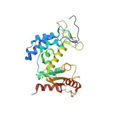
    Nicotinamide adenine dinucleotide (NAD + ) is an essential cofactor participating in a variety of important enzyme-catalyzed physiological and pathophysiological processes. Analogues of NAD + provide key and valuable agents for investigating NAD + -dependent enzymes. In this study, we report the preparation of a novel stable NAD + mimic, 4'-thioribose NAD + (S-NAD + ), using a facile and efficient chemoenzymatic approach. Substrate activity assays indicated the resulting S-NAD + is chemically inert to human CD38 and sirtuin 2 enzymes, but capable of participating in redox reactions in a manner similar to NAD + . X-ray crystallographic analysis revealed binding of S-NAD + to the active site of human CD38 and critical residues involved in leaving group activation and catalysis. By more closely mimicking NAD + in geometry and electrostatics, the generated S-NAD + offers a unique and important tool that can be extended to study enzymes utilizing NAD + .


  • Organizational Affiliation
    • Department of Pharmacology and Pharmaceutical Sciences , School of Pharmacy , University of Southern California , 1985 Zonal Ave , Los Angeles , CA 90089 , USA . Email: yongz@usc.edu.

Macromolecule Content 

  • Total Structure Weight: 60.89 kDa 
  • Atom Count: 4,083 
  • Modeled Residue Count: 495 
  • Deposited Residue Count: 514 
  • Unique protein chains: 1

Macromolecules

Find similar proteins by:|  3D Structure
Entity ID: 1
MoleculeChains  Sequence LengthOrganismDetailsImage
ADP-ribosyl cyclase/cyclic ADP-ribose hydrolase 1
A, B
257Homo sapiensMutation(s): 4 
Gene Names: CD38
EC: 3.2.2.6 (PDB Primary Data), 2.4.99.20 (PDB Primary Data), 3.2.2 (UniProt)
UniProt & NIH Common Fund Data Resources
Find proteins for P28907 (Homo sapiens)
Explore P28907 
Go to UniProtKB:  P28907
PHAROS:  P28907
GTEx:  ENSG00000004468 
Entity Groups
Sequence Clusters30% Identity50% Identity70% Identity90% Identity95% Identity100% Identity
UniProt GroupP28907
Sequence Annotations
Expand
Reference Sequence

Small Molecules

Ligands 1 Unique
IDChains Name / Formula / InChI Key2D Diagram3D Interactions
ZNA
(Subject of Investigation/LOI)

Query on ZNA


Download:Ideal Coordinates CCD File
C [auth A],
D [auth B]
[[(2~{R},3~{S},4~{R},5~{R})-5-(3-aminocarbonylpyridin-1-ium-1-yl)-3,4-bis(oxidanyl)thiolan-2-yl]methoxy-oxidanyl-phosphoryl] [(2~{R},3~{S},4~{R},5~{R})-5-(6-aminopurin-9-yl)-3,4-bis(oxidanyl)oxolan-2-yl]methyl hydrogen phosphate
C21 H28 N7 O13 P2 S
YLEHCOOOOOOSDX-NNYOXOHSSA-O

Experimental Data & Validation

Experimental Data

  • Method: X-RAY DIFFRACTION
  • Resolution: 2.40 Å
  • R-Value Free:  0.262 (Depositor), 0.265 (DCC) 
  • R-Value Work:  0.216 (Depositor), 0.220 (DCC) 
  • R-Value Observed: 0.218 (Depositor) 
Space Group: P 1 21 1
Unit Cell:
Length ( Å )Angle ( ˚ )
a = 57.742α = 90
b = 51.106β = 96.89
c = 100.674γ = 90
Software Package:
Software NamePurpose
REFMACrefinement
XDSdata reduction
SCALAdata scaling
PHASERphasing
PDB_EXTRACTdata extraction

Structure Validation

View Full Validation Report


Ligand Structure Quality Assessment 


Entry History 

& Funding Information

Deposition Data


Funding OrganizationLocationGrant Number
Other privateUnited StatesV2016-021
National Institutes of Health/National Institute of Diabetes and Digestive and Kidney Disease (NIH/NIDDK)United StatesP30DK048522

Revision History  (Full details and data files)

  • Version 1.0: 2018-11-21
    Type: Initial release
  • Version 1.1: 2019-11-13
    Changes: Database references
  • Version 1.2: 2019-12-25
    Changes: Author supporting evidence
  • Version 1.3: 2023-10-11
    Changes: Data collection, Database references, Refinement description
  • Version 2.0: 2024-05-15
    Changes: Derived calculations, Non-polymer description, Structure summary
  • Version 2.1: 2024-11-20
    Changes: Structure summary