6E6L | pdb_00006e6l

Crystal structure of the holo retinal-bound domain-swapped dimer Q108K:K40L:T51F:Y60A mutant of human cellular retinol binding protein II


Experimental Data Snapshot

  • Method: X-RAY DIFFRACTION
  • Resolution: 2.08 Å
  • R-Value Free: 
    0.248 (Depositor), 0.232 (DCC) 
  • R-Value Work: 
    0.198 (Depositor) 
  • R-Value Observed: 
    0.203 (Depositor) 

Starting Model: experimental
View more details

wwPDB Validation 3D Report Full Report

Validation slider image for 6E6L

Ligand Structure Quality Assessment 


This is version 1.4 of the entry. See complete history

Literature

Engineering the hCRBPII Domain-Swapped Dimer into a New Class of Protein Switches.

Ghanbarpour, A.Pinger, C.Esmatpour Salmani, R.Assar, Z.Santos, E.M.Nosrati, M.Pawlowski, K.Spence, D.Vasileiou, C.Jin, X.Borhan, B.Geiger, J.H.

(2019) J Am Chem Soc 141: 17125-17132

  • DOI: https://doi.org/10.1021/jacs.9b04664
  • Primary Citation Related Structures: 
    6E50, 6E51, 6E5E, 6E5Q, 6E5R, 6E5S, 6E6L, 6E7M, 6MCU, 6MCV, 6MKV, 6MLB, 6ON5, 6ON7, 6ON8

  • PubMed Abstract: 

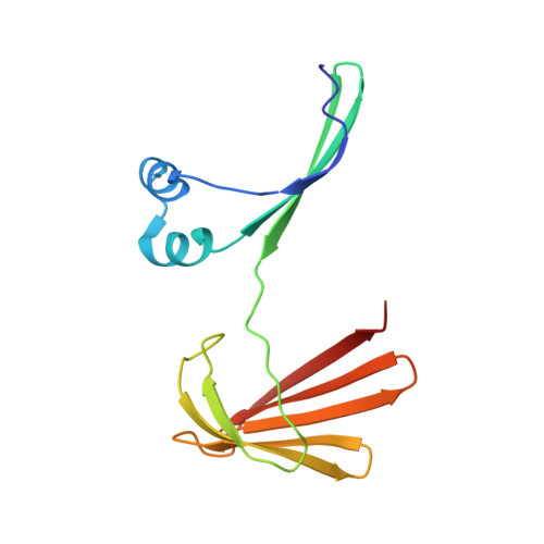
    Protein conformational switches or allosteric proteins play a key role in the regulation of many essential biological pathways. Nonetheless, the implementation of protein conformational switches in protein design applications has proven challenging, with only a few known examples that are not derivatives of naturally occurring allosteric systems. We have discovered that the domain-swapped (DS) dimer of hCRBPII undergoes a large and robust conformational change upon retinal binding, making it a potentially powerful template for the design of protein conformational switches. Atomic resolution structures of the apo- and holo-forms illuminate a simple, mechanical movement involving sterically driven torsion angle flipping of two residues that drive the motion. We further demonstrate that the conformational "readout" can be altered by addition of cross-domain disulfide bonds, also visualized at atomic resolution. Finally, as a proof of principle, we have created an allosteric metal binding site in the DS dimer, where ligand binding results in a reversible 5-fold loss of metal binding affinity. The high resolution structure of the metal-bound variant illustrates a well-formed metal binding site at the interface of the two domains of the DS dimer and confirms the design strategy for allosteric regulation.


  • Organizational Affiliation
    • Department of Chemistry , Michigan State University , East Lansing , Michigan 48824 , United States.

Macromolecule Content 

  • Total Structure Weight: 15.88 kDa 
  • Atom Count: 1,152 
  • Modeled Residue Count: 133 
  • Deposited Residue Count: 133 
  • Unique protein chains: 1

Macromolecules

Find similar proteins by:|  3D Structure
Entity ID: 1
MoleculeChains  Sequence LengthOrganismDetailsImage
Retinol-binding protein 2133Homo sapiensMutation(s): 4 
Gene Names: RBP2CRBP2
UniProt & NIH Common Fund Data Resources
Find proteins for P50120 (Homo sapiens)
Explore P50120 
Go to UniProtKB:  P50120
PHAROS:  P50120
GTEx:  ENSG00000114113 
Entity Groups
Sequence Clusters30% Identity50% Identity70% Identity90% Identity95% Identity100% Identity
UniProt GroupP50120
Sequence Annotations
Expand
Reference Sequence

Experimental Data & Validation

Experimental Data

  • Method: X-RAY DIFFRACTION
  • Resolution: 2.08 Å
  • R-Value Free:  0.248 (Depositor), 0.232 (DCC) 
  • R-Value Work:  0.198 (Depositor) 
  • R-Value Observed: 0.203 (Depositor) 
Space Group: P 21 21 2
Unit Cell:
Length ( Å )Angle ( ˚ )
a = 63.765α = 90
b = 60.554β = 90
c = 36.573γ = 90
Software Package:
Software NamePurpose
PHENIXrefinement
HKL-2000data reduction
HKL-2000data scaling
PHASERphasing

Structure Validation

View Full Validation Report


Ligand Structure Quality Assessment 


Entry History 

& Funding Information

Deposition Data


Funding OrganizationLocationGrant Number
National Institutes of Health/National Human Genome Research Institute (NIH/NHGRI)United States--

Revision History  (Full details and data files)

  • Version 1.0: 2019-10-16
    Type: Initial release
  • Version 1.1: 2019-11-13
    Changes: Data collection, Database references
  • Version 1.2: 2019-12-18
    Changes: Author supporting evidence
  • Version 1.3: 2023-10-11
    Changes: Data collection, Database references, Refinement description
  • Version 1.4: 2024-11-06
    Changes: Structure summary