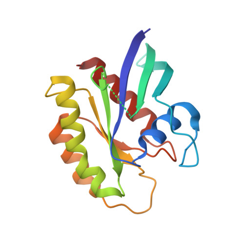
6E6H

NRAS G13D bound to GppNHp (N13GNP)


Experimental Data Snapshot

  • Method: X-RAY DIFFRACTION
  • Resolution: 1.99 Å
  • R-Value Free: 0.236 
  • R-Value Work: 0.179 

wwPDB Validation   3D Report Full Report


Ligand Structure Quality Assessment 


This is version 1.3 of the entry. See complete history


Literature

Isoform-Specific Destabilization of the Active Site Reveals a Molecular Mechanism of Intrinsic Activation of KRas G13D.

Johnson, C.W.Lin, Y.J.Reid, D.Parker, J.Pavlopoulos, S.Dischinger, P.Graveel, C.Aguirre, A.J.Steensma, M.Haigis, K.M.Mattos, C.

(2019) Cell Rep 28: 1538-1550.e7

  • DOI: https://doi.org/10.1016/j.celrep.2019.07.026
  • Primary Citation of Related Structures:  
    6DZH, 6E6C, 6E6F, 6E6G, 6E6H, 6E6P

  • PubMed Abstract: 

    Ras GTPases are mutated at codons 12, 13, and 61, with different frequencies in KRas, HRas, and NRas and in a cancer-specific manner. The G13D mutant appears in 25% of KRas-driven colorectal cancers, while observed only rarely in HRas or NRas. Structures of Ras G13D in the three isoforms show an open active site, with adjustments to the D13 backbone torsion angles and with disconnected switch regions. KRas G13D has unique features that destabilize the nucleotide-binding pocket. In KRas G13D bound to GDP, A59 is placed in the Mg 2+ binding site, as in the HRas-SOS complex. Structure and biochemistry are consistent with an intermediate level of KRas G13D bound to GTP, relative to wild-type and KRas G12D, observed in genetically engineered mouse models. The results explain in part the elevated frequency of the G13D mutant in KRas over the other isoforms of Ras.


  • Organizational Affiliation

    Department of Chemistry and Chemical Biology, Northeastern University, Boston, MA 02115, USA.


Macromolecules
Find similar proteins by:  (by identity cutoff)  |  3D Structure
Entity ID: 1
MoleculeChains Sequence LengthOrganismDetailsImage
GTPase NRas166Homo sapiensMutation(s): 1 
Gene Names: NRASHRAS1
EC: 3.6.5.2
UniProt & NIH Common Fund Data Resources
Find proteins for P01111 (Homo sapiens)
Explore P01111 
Go to UniProtKB:  P01111
PHAROS:  P01111
GTEx:  ENSG00000213281 
Entity Groups  
Sequence Clusters30% Identity50% Identity70% Identity90% Identity95% Identity100% Identity
UniProt GroupP01111
Sequence Annotations
Expand
  • Reference Sequence
Small Molecules
Ligands 2 Unique
IDChains Name / Formula / InChI Key2D Diagram3D Interactions
GNP
Query on GNP

Download Ideal Coordinates CCD File 
C [auth A]PHOSPHOAMINOPHOSPHONIC ACID-GUANYLATE ESTER
C10 H17 N6 O13 P3
UQABYHGXWYXDTK-UUOKFMHZSA-N
MG
Query on MG

Download Ideal Coordinates CCD File 
B [auth A]MAGNESIUM ION
Mg
JLVVSXFLKOJNIY-UHFFFAOYSA-N
Experimental Data & Validation

Experimental Data

  • Method: X-RAY DIFFRACTION
  • Resolution: 1.99 Å
  • R-Value Free: 0.236 
  • R-Value Work: 0.179 
  • Space Group: C 1 2 1
Unit Cell:
Length ( Å )Angle ( ˚ )
a = 59.553α = 90
b = 39.565β = 106.4
c = 65.016γ = 90
Software Package:
Software NamePurpose
HKL-2000data scaling
PHENIXrefinement
PDB_EXTRACTdata extraction
HKL-3000data reduction
PHENIXphasing

Structure Validation

View Full Validation Report


Ligand Structure Quality Assessment 


Entry History & Funding Information

Deposition Data


Funding OrganizationLocationGrant Number
National Science Foundation (NSF, United States)United StatesMCB-1517295

Revision History  (Full details and data files)

  • Version 1.0: 2019-07-31
    Type: Initial release
  • Version 1.1: 2019-09-04
    Changes: Data collection, Database references, Refinement description, Structure summary
  • Version 1.2: 2019-11-27
    Changes: Author supporting evidence
  • Version 1.3: 2024-03-13
    Changes: Data collection, Database references, Refinement description