6E5V | pdb_00006e5v

human mGlu8 receptor amino terminal domain in complex with (S)-3,4-Dicarboxyphenylglycine (DCPG)


Experimental Data Snapshot

  • Method: X-RAY DIFFRACTION
  • Resolution: 2.95 Å
  • R-Value Free: 
    0.306 (Depositor), 0.293 (DCC) 
  • R-Value Work: 
    0.255 (Depositor) 
  • R-Value Observed: 
    0.258 (Depositor) 

Starting Model: experimental
View more details

wwPDB Validation 3D Report Full Report

Validation slider image for 6E5V

Ligand Structure Quality Assessment 


This is version 1.3 of the entry. See complete history

Literature

Structural Basis for ( S)-3,4-Dicarboxyphenylglycine (DCPG) As a Potent and Subtype Selective Agonist of the mGlu8Receptor.

Chen, Q.Ho, J.D.Ashok, S.Vargas, M.C.Wang, J.Atwell, S.Bures, M.Schkeryantz, J.M.Monn, J.A.Hao, J.

(2018) J Med Chem 61: 10040-10052

  • DOI: https://doi.org/10.1021/acs.jmedchem.8b01120
  • Primary Citation Related Structures: 
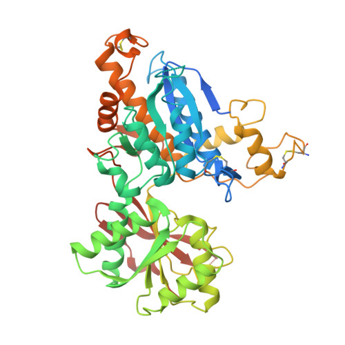
    6E5V

  • PubMed Abstract: 

    ( S)-3,4-Dicarboxyphenylglycine (DCPG) was first reported in 2001 as a potent orthosteric agonist with high subtype selectivity for the mGlu 8 receptor, but the structural basis for its high selectivity is not well understood. We have solved a cocrystal structure of recombinant human mGlu 8 amino terminal domain (ATD) protein bound to ( S)-DCPG, which possesses the largest lobe opening angle observed to date among known agonist-bound mGlu ATD crystal structures. The binding conformation of ( S)-DCPG observed in the crystal structure is significantly different from that in the homology model built from an l-glutamate-bound rat mGlu 1 ATD crystal structure, which has a smaller lobe opening angle. This highlights the importance of considering various lobe opening angles when modeling mGlu ATD-ligand complex. New homology models of other mGlu receptors based on the ( S)-DCPG-bound mGlu 8 ATD crystal structure were explored to rationalize ( S)-DCPG's high mGlu 8 receptor subtype selectivity.

Macromolecule Content 

  • Total Structure Weight: 118.21 kDa 
  • Atom Count: 6,754 
  • Modeled Residue Count: 882 
  • Deposited Residue Count: 1,046 
  • Unique protein chains: 1

Macromolecules

Find similar proteins by:|  3D Structure
Entity ID: 1
MoleculeChains  Sequence LengthOrganismDetailsImage
Metabotropic glutamate receptor 8
A, B
523Homo sapiensMutation(s): 1 
Gene Names: GRM8GPRC1HMGLUR8
UniProt & NIH Common Fund Data Resources
Find proteins for O00222 (Homo sapiens)
Explore O00222 
Go to UniProtKB:  O00222
PHAROS:  O00222
GTEx:  ENSG00000179603 
Entity Groups
Sequence Clusters30% Identity50% Identity70% Identity90% Identity95% Identity100% Identity
UniProt GroupO00222
Sequence Annotations
Expand
Reference Sequence

Experimental Data & Validation

Experimental Data

  • Method: X-RAY DIFFRACTION
  • Resolution: 2.95 Å
  • R-Value Free:  0.306 (Depositor), 0.293 (DCC) 
  • R-Value Work:  0.255 (Depositor) 
  • R-Value Observed: 0.258 (Depositor) 
Space Group: P 21 21 21
Unit Cell:
Length ( Å )Angle ( ˚ )
a = 82.945α = 90
b = 106.78β = 90
c = 117.908γ = 90
Software Package:
Software NamePurpose
REFMACrefinement
PDB_EXTRACTdata extraction
MOSFLMdata reduction
SCALAdata scaling
PHASERphasing

Structure Validation

View Full Validation Report


Ligand Structure Quality Assessment 


Entry History 

Deposition Data

Revision History  (Full details and data files)

  • Version 1.0: 2018-11-07
    Type: Initial release
  • Version 1.1: 2018-12-05
    Changes: Data collection, Database references
  • Version 1.2: 2023-10-11
    Changes: Data collection, Database references, Refinement description
  • Version 1.3: 2024-11-20
    Changes: Structure summary