6E3B | pdb_00006e3b

STRUCTURE OF Siw14 CATALYTIC CORE

  • Classification: HYDROLASE
  • Organism(s): Saccharomyces cerevisiae
  • Expression System: Escherichia coli
  • Mutation(s): No 

  • Deposited: 2018-07-13 Released: 2019-02-27 
  • Deposition Author(s): Florio, T., Lokareddy, R., Cingolani, G.
  • Funding Organization(s): National Institutes of Health/National Cancer Institute (NIH/NCI), National Institutes of Health/National Institute of General Medical Sciences (NIH/NIGMS), National Institutes of Health/Office of the Director

Experimental Data Snapshot

  • Method: X-RAY DIFFRACTION
  • Resolution: 2.50 Å
  • R-Value Free: 
    0.246 (Depositor), 0.216 (DCC) 
  • R-Value Work: 
    0.230 (Depositor), 0.210 (DCC) 
  • R-Value Observed: 
    0.243 (Depositor) 

Starting Model: experimental
View more details

wwPDB Validation 3D Report Full Report

Validation slider image for 6E3B

This is version 1.2 of the entry. See complete history

Literature

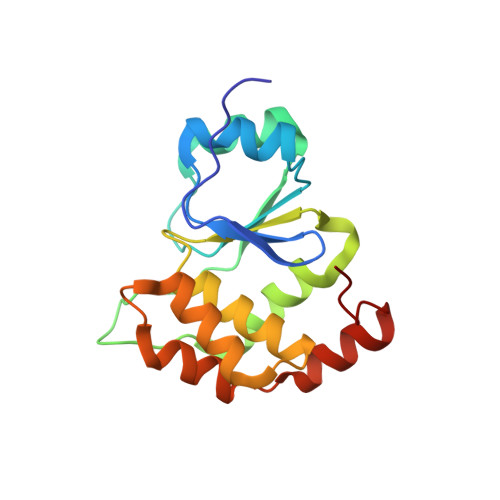
Molecular Architecture of the Inositol Phosphatase Siw14.

Florio, T.J.Lokareddy, R.K.Gillilan, R.E.Cingolani, G.

(2019) Biochemistry 58: 534-545

  • DOI: https://doi.org/10.1021/acs.biochem.8b01044
  • Primary Citation Related Structures: 
    6E3B

  • PubMed Abstract: 

    Siw14 is a recently discovered inositol phosphatase implicated in suppressing prion propagation in Saccharomyces cerevisiae. In this paper, we used hybrid structural methods to decipher Siw14 molecular architecture. We found the protein exists in solution as an elongated monomer that is ∼140 Å in length, containing an acidic N-terminal domain and a basic C-terminal dual-specificity phosphatase (DSP) domain, structurally similar to the glycogen phosphatase laforin. The two domains are connected by a protease susceptible linker and do not interact in vitro. The crystal structure of Siw14-DSP reveals a highly basic phosphate-binding loop and an ∼10 Å deep substrate-binding crevice that evolved to dephosphorylate pyro-phosphate moieties. A pseudoatomic model of the full-length phosphatase generated from solution, crystallographic, biochemical, and modeling data sheds light on the interesting zwitterionic nature of Siw14, which we hypothesized may play a role in discriminating negatively charged inositol phosphates.


  • Organizational Affiliation
    • Department of Biochemistry and Molecular Biology , Thomas Jefferson University , 233 South 10th Street , Philadelphia , Pennsylvania 19107 , United States.

Macromolecule Content 

  • Total Structure Weight: 479.02 kDa 
  • Atom Count: 34,047 
  • Modeled Residue Count: 4,055 
  • Deposited Residue Count: 4,056 
  • Unique protein chains: 1

Macromolecules

Find similar proteins by:|  3D Structure
Entity ID: 1
MoleculeChains  Sequence LengthOrganismDetailsImage
Tyrosine-protein phosphatase SIW14169Saccharomyces cerevisiaeMutation(s): 0 
Gene Names: SIW14
EC: 3.1.3.48 (PDB Primary Data), 3.6.1.52 (UniProt)
UniProt
Find proteins for P53965 (Saccharomyces cerevisiae (strain ATCC 204508 / S288c))
Explore P53965 
Go to UniProtKB:  P53965
Entity Groups
Sequence Clusters30% Identity50% Identity70% Identity90% Identity95% Identity100% Identity
UniProt GroupP53965
Sequence Annotations
Expand
Reference Sequence

Small Molecules

Ligands 1 Unique
IDChains Name / Formula / InChI Key2D Diagram3D Interactions
SO4

Query on SO4


Download:Ideal Coordinates CCD File
AA [auth C]
BA [auth D]
CA [auth E]
DA [auth F]
EA [auth G]
AA [auth C],
BA [auth D],
CA [auth E],
DA [auth F],
EA [auth G],
FA [auth H],
GA [auth I],
HA [auth J],
IA [auth K],
JA [auth L],
KA [auth M],
LA [auth N],
MA [auth O],
NA [auth P],
OA [auth Q],
PA [auth R],
QA [auth S],
RA [auth T],
SA [auth V],
TA [auth W],
UA [auth X],
VA [auth Y],
Y [auth A],
Z [auth B]
SULFATE ION
O4 S
QAOWNCQODCNURD-UHFFFAOYSA-L

Experimental Data & Validation

Experimental Data

  • Method: X-RAY DIFFRACTION
  • Resolution: 2.50 Å
  • R-Value Free:  0.246 (Depositor), 0.216 (DCC) 
  • R-Value Work:  0.230 (Depositor), 0.210 (DCC) 
  • R-Value Observed: 0.243 (Depositor) 
Space Group: C 1 2 1
Unit Cell:
Length ( Å )Angle ( ˚ )
a = 92.467α = 90
b = 160.487β = 90.32
c = 360.572γ = 90
Software Package:
Software NamePurpose
PHENIXrefinement
PDB_EXTRACTdata extraction
HKL-2000data reduction
HKL-2000data scaling
PHASERphasing

Structure Validation

View Full Validation Report


Entry History 

& Funding Information

Deposition Data


Funding OrganizationLocationGrant Number
National Institutes of Health/National Cancer Institute (NIH/NCI)United StatesP30CA56036
National Institutes of Health/National Institute of General Medical Sciences (NIH/NIGMS)United StatesR01GM122844
National Institutes of Health/Office of the DirectorUnited StatesS10OD017987

Revision History  (Full details and data files)

  • Version 1.0: 2019-02-27
    Type: Initial release
  • Version 1.1: 2019-12-04
    Changes: Author supporting evidence
  • Version 1.2: 2023-10-11
    Changes: Data collection, Database references, Refinement description