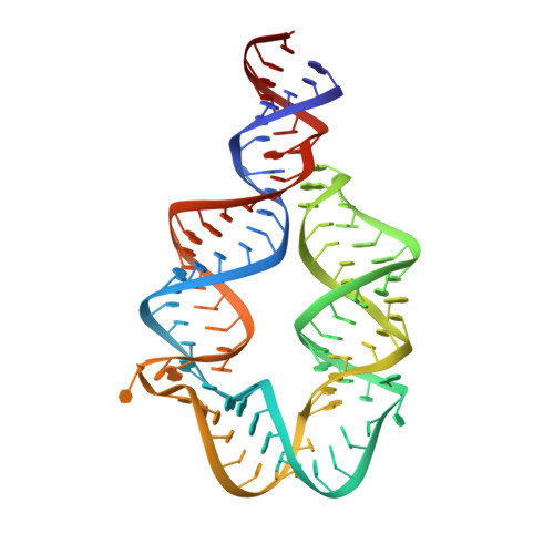
6DVK

Computationally designed mini tetraloop-tetraloop receptor by the RNAMake program - construct 6 (miniTTR 6)


Experimental Data Snapshot

  • Method: X-RAY DIFFRACTION
  • Resolution: 2.55 Å
  • R-Value Free: 0.269 
  • R-Value Work: 0.232 
  • R-Value Observed: 0.233 

wwPDB Validation   3D Report Full Report


This is version 1.5 of the entry. See complete history


Literature

Computational design of three-dimensional RNA structure and function.

Yesselman, J.D.Eiler, D.Carlson, E.D.Gotrik, M.R.d'Aquino, A.E.Ooms, A.N.Kladwang, W.Carlson, P.D.Shi, X.Costantino, D.A.Herschlag, D.Lucks, J.B.Jewett, M.C.Kieft, J.S.Das, R.

(2019) Nat Nanotechnol 14: 866-873

  • DOI: https://doi.org/10.1038/s41565-019-0517-8
  • Primary Citation of Related Structures:  
    6DVK

  • PubMed Abstract: 

    RNA nanotechnology seeks to create nanoscale machines by repurposing natural RNA modules. The field is slowed by the current need for human intuition during three-dimensional structural design. Here, we demonstrate that three distinct problems in RNA nanotechnology can be reduced to a pathfinding problem and automatically solved through an algorithm called RNAMake. First, RNAMake discovers highly stable single-chain solutions to the classic problem of aligning a tetraloop and its sequence-distal receptor, with experimental validation from chemical mapping, gel electrophoresis, solution X-ray scattering and crystallography with 2.55 Å resolution. Second, RNAMake automatically generates structured tethers that integrate 16S and 23S ribosomal RNAs into single-chain ribosomal RNAs that remain uncleaved by ribonucleases and assemble onto messenger RNA. Third, RNAMake enables the automated stabilization of small-molecule binding RNAs, with designed tertiary contacts that improve the binding affinity of the ATP aptamer and improve the fluorescence and stability of the Spinach RNA in cell extracts and in living Escherichia coli cells.


  • Organizational Affiliation

    Department of Biochemistry, Stanford University School of Medicine, Stanford, CA, USA.


Macromolecules
Find similar nucleic acids by:  (by identity cutoff)  |  3D Structure
Entity ID: 1
MoleculeChains LengthOrganismImage
RNA (95-MER)A [auth H]95synthetic construct
Sequence Annotations
Expand
  • Reference Sequence
Small Molecules
Ligands 2 Unique
IDChains Name / Formula / InChI Key2D Diagram3D Interactions
CO
Query on CO

Download Ideal Coordinates CCD File 
F [auth H],
G [auth H],
H,
I [auth H]
COBALT (II) ION
Co
XLJKHNWPARRRJB-UHFFFAOYSA-N
MG
Query on MG

Download Ideal Coordinates CCD File 
B [auth H],
C [auth H],
D [auth H],
E [auth H],
J [auth H]
MAGNESIUM ION
Mg
JLVVSXFLKOJNIY-UHFFFAOYSA-N
Experimental Data & Validation

Experimental Data

  • Method: X-RAY DIFFRACTION
  • Resolution: 2.55 Å
  • R-Value Free: 0.269 
  • R-Value Work: 0.232 
  • R-Value Observed: 0.233 
  • Space Group: C 1 2 1
Unit Cell:
Length ( Å )Angle ( ˚ )
a = 233.372α = 90
b = 25.358β = 99.74
c = 42.861γ = 90
Software Package:
Software NamePurpose
REFMACrefinement
XSCALEdata scaling
PDB_EXTRACTdata extraction
XDSdata reduction
PHASERphasing

Structure Validation

View Full Validation Report


Entry History & Funding Information

Deposition Data


Funding OrganizationLocationGrant Number
National Institutes of Health/National Institute of General Medical Sciences (NIH/NIGMS)United StatesR35GM118070
National Institutes of Health/National Institute of General Medical Sciences (NIH/NIGMS)United StatesGM100953
National Institutes of Health/National Institute of General Medical Sciences (NIH/NIGMS)United StatesGM112294

Revision History  (Full details and data files)

  • Version 1.0: 2019-06-26
    Type: Initial release
  • Version 1.1: 2019-09-04
    Changes: Data collection, Database references
  • Version 1.2: 2019-09-18
    Changes: Data collection, Database references
  • Version 1.3: 2020-01-01
    Changes: Author supporting evidence
  • Version 1.4: 2020-01-15
    Changes: Data collection
  • Version 1.5: 2024-03-13
    Changes: Data collection, Database references, Derived calculations