


Role of Bound Water Molecules in Differential Recognition of Di-Acetylated Histone Peptides by the BRD4 Bromodomains
Babault, N., Zhang, Q., Zhang, G., Zhou, M.M., Zeng, L.To be published.
Experimental Data Snapshot
Entity ID: 1 | |||||
|---|---|---|---|---|---|
| Molecule | Chains | Sequence Length | Organism | Details | Image |
| Bromodomain-containing protein 4 | 112 | Homo sapiens | Mutation(s): 0 Gene Names: BRD4, HUNK1 | 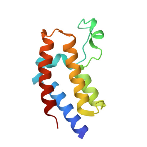 | |
UniProt & NIH Common Fund Data Resources | |||||
PHAROS: O60885 GTEx: ENSG00000141867 | |||||
Entity Groups | |||||
| Sequence Clusters | 30% Identity50% Identity70% Identity90% Identity95% Identity100% Identity | ||||
| UniProt Group | O60885 | ||||
Sequence AnnotationsExpand | |||||
Reference Sequence | |||||
| Ligands 2 Unique | |||||
|---|---|---|---|---|---|
| ID | Chains | Name / Formula / InChI Key | 2D Diagram | 3D Interactions | |
| 0S6 Download:Ideal Coordinates CCD File | C [auth A], D [auth A], G [auth B] | methyl [(6S)-4-(4-chlorophenyl)-2,3,9-trimethyl-6H-thieno[3,2-f][1,2,4]triazolo[4,3-a][1,4]diazepin-6-yl]acetate C20 H19 Cl N4 O2 S GGRCIHACOIMRKY-HNNXBMFYSA-N |  | ||
| EDO Download:Ideal Coordinates CCD File | E [auth A] F [auth A] H [auth B] I [auth B] J [auth B] | 1,2-ETHANEDIOL C2 H6 O2 LYCAIKOWRPUZTN-UHFFFAOYSA-N |  | ||
| Length ( Å ) | Angle ( ˚ ) |
|---|---|
| a = 74.58 | α = 90 |
| b = 74.58 | β = 90 |
| c = 111.47 | γ = 120 |
| Software Name | Purpose |
|---|---|
| SCALA | data scaling |
| PHENIX | refinement |
| PDB_EXTRACT | data extraction |
| iMOSFLM | data reduction |
| PHASER | phasing |
| Funding Organization | Location | Grant Number |
|---|---|---|
| National Institutes of Health/National Institute of General Medical Sciences (NIH/NIGMS) | United States | R01HG004508 |


