6DUB | pdb_00006dub

Crystal structure of a methyltransferase


Experimental Data Snapshot

  • Method: X-RAY DIFFRACTION
  • Resolution: 1.20 Å
  • R-Value Free: 
    0.178 (Depositor), 0.184 (DCC) 
  • R-Value Work: 
    0.154 (Depositor), 0.163 (DCC) 
  • R-Value Observed: 
    0.155 (Depositor) 

Starting Model: experimental
View more details

wwPDB Validation 3D Report Full Report

Validation slider image for 6DUB

Ligand Structure Quality Assessment 


This is version 1.2 of the entry. See complete history

Literature

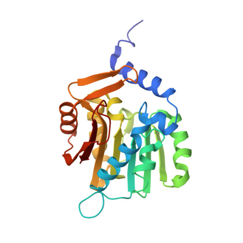
An asparagine/glycine switch governs product specificity of human N-terminal methyltransferase NTMT2.

Dong, C.Dong, G.Li, L.Zhu, L.Tempel, W.Liu, Y.Huang, R.Min, J.

(2018) Commun Biol 1: 183-183

  • DOI: https://doi.org/10.1038/s42003-018-0196-2
  • Primary Citation Related Structures: 
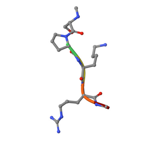
    5UBB, 6DUB

  • PubMed Abstract: 

    α-N-terminal methylation of proteins is an important post-translational modification that is catalyzed by two different N-terminal methyltransferases, namely NTMT1 and NTMT2. Previous studies have suggested that NTMT1 is a tri-methyltransferase, whereas NTMT2 is a mono-methyltransferase. Here, we report the first crystal structures, to our knowledge, of NTMT2 in binary complex with S-adenosyl-L-methionine as well as in ternary complex with S-adenosyl-L-homocysteine and a substrate peptide. Our structural observations combined with biochemical studies reveal that NTMT2 is also able to di-/tri-methylate the GPKRIA peptide and di-methylate the PPKRIA peptide, otherwise it is predominantly a mono-methyltransferase. The residue N89 of NTMT2 serves as a gatekeeper residue that regulates the binding of unmethylated versus monomethylated substrate peptide. Structural comparison of NTMT1 and NTMT2 prompts us to design a N89G mutant of NTMT2 that can profoundly alter its catalytic activities and product specificities.


  • Organizational Affiliation
    • Structural Genomics Consortium, University of Toronto, Toronto, M5G1L7, ON, Canada.

Macromolecule Content 

  • Total Structure Weight: 52.43 kDa 
  • Atom Count: 4,160 
  • Modeled Residue Count: 450 
  • Deposited Residue Count: 456 
  • Unique protein chains: 2

Macromolecules

Find similar proteins by:|  3D Structure
Entity ID: 1
MoleculeChains  Sequence LengthOrganismDetailsImage
Alpha N-terminal protein methyltransferase 1B
A, B
222Homo sapiensMutation(s): 0 
Gene Names: METTL11BC1orf184NRMT2
EC: 2.1.1.299
UniProt & NIH Common Fund Data Resources
Find proteins for Q5VVY1 (Homo sapiens)
Explore Q5VVY1 
Go to UniProtKB:  Q5VVY1
GTEx:  ENSG00000203740 
Entity Groups
Sequence Clusters30% Identity50% Identity70% Identity90% Identity95% Identity100% Identity
UniProt GroupQ5VVY1
Sequence Annotations
Expand
Reference Sequence
Find similar proteins by:  Sequence   |   3D Structure  
Entity ID: 2
MoleculeChains  Sequence LengthOrganismDetailsImage
RCC1C [auth E],
D [auth F]
6Homo sapiensMutation(s): 0 
UniProt & NIH Common Fund Data Resources
Find proteins for P18754 (Homo sapiens)
Explore P18754 
Go to UniProtKB:  P18754
PHAROS:  P18754
GTEx:  ENSG00000180198 
Entity Groups
UniProt GroupP18754
Sequence Annotations
Expand
Reference Sequence

Small Molecules

Ligands 3 Unique
IDChains Name / Formula / InChI Key2D Diagram3D Interactions
SAH

Query on SAH


Download:Ideal Coordinates CCD File
E [auth A],
PA [auth B]
S-ADENOSYL-L-HOMOCYSTEINE
C14 H20 N6 O5 S
ZJUKTBDSGOFHSH-WFMPWKQPSA-N
GOL

Query on GOL


Download:Ideal Coordinates CCD File
F [auth A],
G [auth A],
QA [auth B]
GLYCEROL
C3 H8 O3
PEDCQBHIVMGVHV-UHFFFAOYSA-N
UNX

Query on UNX


Download:Ideal Coordinates CCD File
AA [auth A]
AB [auth B]
BA [auth A]
BB [auth B]
CA [auth A]
AA [auth A],
AB [auth B],
BA [auth A],
BB [auth B],
CA [auth A],
CB [auth B],
DA [auth A],
DB [auth B],
EA [auth A],
EB [auth B],
FA [auth A],
FB [auth B],
GA [auth A],
GB [auth B],
H [auth A],
HA [auth A],
HB [auth B],
I [auth A],
IA [auth A],
IB [auth B],
J [auth A],
JA [auth A],
JB [auth B],
K [auth A],
KA [auth A],
KB [auth B],
L [auth A],
LA [auth A],
LB [auth B],
M [auth A],
MA [auth A],
MB [auth B],
N [auth A],
NA [auth A],
NB [auth E],
O [auth A],
OA [auth A],
OB [auth E],
P [auth A],
PB [auth E],
Q [auth A],
QB [auth F],
R [auth A],
RA [auth B],
S [auth A],
SA [auth B],
T [auth A],
TA [auth B],
U [auth A],
UA [auth B],
V [auth A],
VA [auth B],
W [auth A],
WA [auth B],
X [auth A],
XA [auth B],
Y [auth A],
YA [auth B],
Z [auth A],
ZA [auth B]
UNKNOWN ATOM OR ION
X

Experimental Data & Validation

Experimental Data

  • Method: X-RAY DIFFRACTION
  • Resolution: 1.20 Å
  • R-Value Free:  0.178 (Depositor), 0.184 (DCC) 
  • R-Value Work:  0.154 (Depositor), 0.163 (DCC) 
  • R-Value Observed: 0.155 (Depositor) 
Space Group: P 43
Unit Cell:
Length ( Å )Angle ( ˚ )
a = 44.359α = 90
b = 44.359β = 90
c = 262.058γ = 90
Software Package:
Software NamePurpose
REFMACrefinement
Aimlessdata scaling
PDB_EXTRACTdata extraction
HKL-3000data reduction
PHASERphasing

Structure Validation

View Full Validation Report


Ligand Structure Quality Assessment 


Entry History 

Deposition Data

Revision History  (Full details and data files)

  • Version 1.0: 2018-07-25
    Type: Initial release
  • Version 1.1: 2018-11-28
    Changes: Data collection, Database references
  • Version 1.2: 2023-10-11
    Changes: Data collection, Database references, Refinement description