Clarifying the Copper Coordination Environment in a de Novo Designed Red Copper Protein.
Koebke, K.J., Ruckthong, L., Meagher, J.L., Mathieu, E., Harland, J., Deb, A., Lehnert, N., Policar, C., Tard, C., Penner-Hahn, J.E., Stuckey, J.A., Pecoraro, V.L.(2018) Inorg Chem 57: 12291-12302
- PubMed: 30226758 Search on PubMedSearch on PubMed Central
- DOI: https://doi.org/10.1021/acs.inorgchem.8b01989
- Primary Citation Related Structures:
6DS9 - PubMed Abstract:
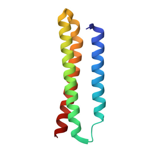
Cupredoxins are copper-dependent electron-transfer proteins that can be categorized as blue, purple, green, and red depending on the spectroscopic properties of the Cu(II) bound forms. Interestingly, despite significantly different first coordination spheres and nuclearity, all cupredoxins share a common Greek Key β-sheet fold. We have previously reported the design of a red copper protein within a completely distinct three-helical bundle protein, α 3 DChC2. (1) While this design demonstrated that a β-barrel fold was not requisite to recapitulate the properties of a native cupredoxin center, the parent peptide α 3 D was not sufficiently stable to allow further study through additional mutations. Here we present the design of an elongated protein GRANDα 3 D (GRα 3 D) with Δ G u = -11.4 kcal/mol compared to the original design's -5.1 kcal/mol. Diffraction quality crystals were grown of GRα 3 D (a first for an α 3 D peptide) and solved to a resolution of 1.34 Å. Examination of this structure suggested that Glu41 might interact with the Cu in our previously reported red copper protein. The previous bis(histidine)(cysteine) site (GRα 3 DChC2) was designed into this new scaffold and a series of variant constructs were made to explore this hypothesis. Mutation studies around Glu41 not only prove the proposed interaction, but also enabled tuning of the constructs' hyperfine coupling constant from 160 to 127 × 10 -4 cm -1 . X-ray absorption spectroscopy analysis is consistent with these hyperfine coupling differences being the result of variant 4p mixing related to coordination geometry changes. These studies not only prove that an Glu41-Cu interaction leads to the α 3 DChC2 construct's red copper protein like spectral properties, but also exemplify the exact control one can have in a de novo construct to tune the properties of an electron-transfer Cu site.
- Laboratoire des biomolécules, LBM, Département de chimie , École normale supérieure, PSL University, Sorbonne Université, CNRS , 75005 Paris , France.
Organizational Affiliation: