6DQY | pdb_00006dqy

Crystal Structure analysis of Superoxide Dismutase from Trichoderma reesei


Experimental Data Snapshot

  • Method: X-RAY DIFFRACTION
  • Resolution: 2.30 Å
  • R-Value Free: 
    0.207 (Depositor), 0.207 (DCC) 
  • R-Value Work: 
    0.162 (Depositor), 0.164 (DCC) 
  • R-Value Observed: 
    0.164 (Depositor) 

wwPDB Validation 3D Report Full Report

Validation slider image for 6DQY

This is version 1.2 of the entry. See complete history

Literature

Crystal Structure of Superoxide Dismutase from Trichoderma reesei

Mendoza, E.R.Stelmastchuk, L.B.F.Ferreira Junior, J.R.S.Garratt, R.C.

To be published.

Macromolecule Content 

  • Total Structure Weight: 559.57 kDa 
  • Atom Count: 41,443 
  • Modeled Residue Count: 4,983 
  • Deposited Residue Count: 5,088 
  • Unique protein chains: 1

Macromolecules

Find similar proteins by:|  3D Structure
Entity ID: 1
MoleculeChains  Sequence LengthOrganismDetailsImage
Superoxide dismutase
A, B, C, D, E
A, B, C, D, E, F, G, H, I, J, K, L, M, N, O, P, Q, R, S, T, U, V, W, X
212Trichoderma reesei QM6aMutation(s): 0 
Gene Names: TRIREDRAFT_66345
EC: 1.15.1.1
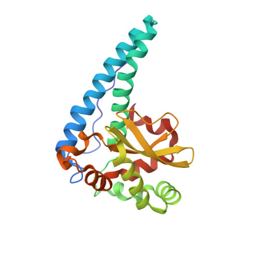
UniProt
Find proteins for G0RQS7 (Hypocrea jecorina (strain QM6a))
Explore G0RQS7 
Go to UniProtKB:  G0RQS7
Entity Groups
Sequence Clusters30% Identity50% Identity70% Identity90% Identity95% Identity100% Identity
UniProt GroupG0RQS7
Sequence Annotations
Expand
Reference Sequence

Small Molecules

Ligands 1 Unique
IDChains Name / Formula / InChI Key2D Diagram3D Interactions
MN

Query on MN


Download:Ideal Coordinates CCD File
AA [auth C]
BA [auth D]
CA [auth E]
DA [auth F]
EA [auth G]
AA [auth C],
BA [auth D],
CA [auth E],
DA [auth F],
EA [auth G],
FA [auth H],
GA [auth I],
HA [auth J],
IA [auth K],
JA [auth L],
KA [auth M],
LA [auth N],
MA [auth O],
NA [auth P],
OA [auth Q],
PA [auth R],
QA [auth S],
RA [auth T],
SA [auth U],
TA [auth V],
UA [auth W],
VA [auth X],
Y [auth A],
Z [auth B]
MANGANESE (II) ION
Mn
WAEMQWOKJMHJLA-UHFFFAOYSA-N

Experimental Data & Validation

Experimental Data

  • Method: X-RAY DIFFRACTION
  • Resolution: 2.30 Å
  • R-Value Free:  0.207 (Depositor), 0.207 (DCC) 
  • R-Value Work:  0.162 (Depositor), 0.164 (DCC) 
  • R-Value Observed: 0.164 (Depositor) 
Space Group: P 1
Unit Cell:
Length ( Å )Angle ( ˚ )
a = 73.77α = 91.59
b = 113.06β = 93.79
c = 162.71γ = 89.98
Software Package:
Software NamePurpose
PHENIXrefinement
SCALAdata scaling
PDB_EXTRACTdata extraction
iMOSFLMdata reduction
PHENIXphasing

Structure Validation

View Full Validation Report


Entry History 

& Funding Information

Deposition Data


Funding OrganizationLocationGrant Number
Sao Paulo Research Foundation (FAPESP)Brazil2014/01855-2

Revision History  (Full details and data files)

  • Version 1.0: 2018-12-12
    Type: Initial release
  • Version 1.1: 2020-01-01
    Changes: Author supporting evidence
  • Version 1.2: 2024-03-13
    Changes: Data collection, Database references, Derived calculations, Refinement description