6DPQ

Mapping the binding trajectory of a suicide inhibitor in human indoleamine 2,3-dioxygenase 1


Experimental Data Snapshot

  • Method: X-RAY DIFFRACTION
  • Resolution: 2.94 Å
  • R-Value Free: 
    0.240 (Depositor), 0.240 (DCC) 
  • R-Value Work: 
    0.191 (Depositor), 0.200 (DCC) 
  • R-Value Observed: 
    0.193 (Depositor) 

Starting Model: experimental
View more details

wwPDB Validation   3D Report Full Report


Ligand Structure Quality Assessment 

Created with Raphaël 2.3.0Worse 01 BetterLigand structure goodness of fit to experimental dataBest fitted HEMClick on this verticalbar to view detailsBest fitted H7PClick on this verticalbar to view details

This is version 1.4 of the entry. See complete history


Literature

Mapping the Binding Trajectory of a Suicide Inhibitor in Human Indoleamine 2,3-Dioxygenase 1.

Pham, K.N.Yeh, S.R.

(2018) J Am Chem Soc 140: 14538-14541

  • DOI: https://doi.org/10.1021/jacs.8b07994
  • Primary Citation of Related Structures:  
    6DPQ, 6DPR, 6MQ6

  • PubMed Abstract: 

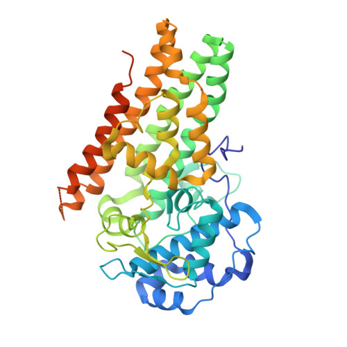
    Human indoleamine 2,3-dioxygenase 1 (hIDO1) is an important heme-containing enzyme that is a key drug target for cancer immunotherapy. Several hIDO1 inhibitors have entered clinical trials, among which BMS-986205 (BMS) stands out as the only suicide inhibitor. Despite its "best-in-class" activity, the action mechanism of BMS remains elusive. Here, we report three crystal structures of hIDO1-BMS complexes that define the complete binding trajectory of the inhibitor. BMS first binds in a solvent exposed surface cleft near the active site in an extended conformation. The initial binding partially unfolds the active site, which triggers heme release, thereby exposing a new binding pocket. The inhibitor then undergoes a large scale movement to this new binding pocket, where it binds by adopting a high energy kinked conformation. Finally, the inhibitor relaxes to a bent conformation, via an additional large scale rearrangement, culminating in the energy minimum state. The structural data offer a molecular explanation for the remarkable efficacy and suicide inhibition activity of the inhibitor. They also suggest a novel strategy that can be applied for drug development targeting hIDO1 and related enzymes.


  • Organizational Affiliation

    Department of Physiology and Biophysics , Albert Einstein College of Medicine , Bronx , New York 10461 , United States.


Macromolecules
Find similar proteins by:  (by identity cutoff)  |  3D Structure
Entity ID: 1
MoleculeChains Sequence LengthOrganismDetailsImage
Indoleamine 2,3-dioxygenase 1
A, B
425Homo sapiensMutation(s): 0 
Gene Names: IDO1IDOINDO
EC: 1.13.11.52
UniProt & NIH Common Fund Data Resources
Find proteins for P14902 (Homo sapiens)
Explore P14902 
Go to UniProtKB:  P14902
PHAROS:  P14902
GTEx:  ENSG00000131203 
Entity Groups  
Sequence Clusters30% Identity50% Identity70% Identity90% Identity95% Identity100% Identity
UniProt GroupP14902
Sequence Annotations
Expand
  • Reference Sequence
Small Molecules
Ligands 3 Unique
IDChains Name / Formula / InChI Key2D Diagram3D Interactions
HEM
Query on HEM

Download Ideal Coordinates CCD File 
C [auth A]PROTOPORPHYRIN IX CONTAINING FE
C34 H32 Fe N4 O4
KABFMIBPWCXCRK-RGGAHWMASA-L
H7P
Query on H7P

Download Ideal Coordinates CCD File 
G [auth B](2R)-N-(4-chlorophenyl)-2-[cis-4-(6-fluoroquinolin-4-yl)cyclohexyl]propanamide
C24 H24 Cl F N2 O
KRTIYQIPSAGSBP-ZACQAIPSSA-N
GOL
Query on GOL

Download Ideal Coordinates CCD File 
D [auth A]
E [auth A]
F [auth A]
H [auth B]
I [auth B]
GLYCEROL
C3 H8 O3
PEDCQBHIVMGVHV-UHFFFAOYSA-N
Binding Affinity Annotations 
IDSourceBinding Affinity
H7P BindingDB:  6DPQ Kd: 470 (nM) from 1 assay(s)
IC50: min: 0.5, max: 1700 (nM) from 11 assay(s)
Experimental Data & Validation

Experimental Data

  • Method: X-RAY DIFFRACTION
  • Resolution: 2.94 Å
  • R-Value Free:  0.240 (Depositor), 0.240 (DCC) 
  • R-Value Work:  0.191 (Depositor), 0.200 (DCC) 
  • R-Value Observed: 0.193 (Depositor) 
Space Group: P 21 21 21
Unit Cell:
Length ( Å )Angle ( ˚ )
a = 85.604α = 90
b = 96.565β = 90
c = 130.764γ = 90
Software Package:
Software NamePurpose
REFMACrefinement
XDSdata reduction
SCALAdata scaling
PHASERphasing

Structure Validation

View Full Validation Report


Ligand Structure Quality Assessment 

Created with Raphaël 2.3.0Worse 01 BetterLigand structure goodness of fit to experimental dataBest fitted HEMClick on this verticalbar to view detailsBest fitted H7PClick on this verticalbar to view details

Entry History & Funding Information

Deposition Data


Funding OrganizationLocationGrant Number
National Institutes of Health/National Institute of General Medical Sciences (NIH/NIGMS)United StatesGM115773
National Science Foundation (NSF, United States)United StatesCHE-1404929

Revision History  (Full details and data files)

  • Version 1.0: 2018-11-07
    Type: Initial release
  • Version 1.1: 2018-11-21
    Changes: Data collection, Database references
  • Version 1.2: 2019-11-27
    Changes: Author supporting evidence
  • Version 1.3: 2023-10-11
    Changes: Data collection, Database references, Refinement description
  • Version 1.4: 2024-11-06
    Changes: Structure summary