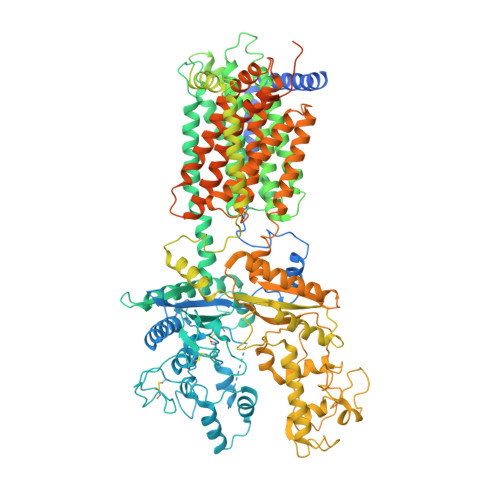
6DMO

Cryo-EM structure of human Ptch1 with three mutations L282Q/T500F/P504L


Experimental Data Snapshot

  • Method: ELECTRON MICROSCOPY
  • Resolution: 4.10 Å
  • Aggregation State: PARTICLE 
  • Reconstruction Method: SINGLE PARTICLE 

wwPDB Validation   3D Report Full Report


This is version 2.1 of the entry. See complete history


Literature

Structural basis for the recognition of Sonic Hedgehog by human Patched1.

Gong, X.Qian, H.Cao, P.Zhao, X.Zhou, Q.Lei, J.Yan, N.

(2018) Science 361

  • DOI: https://doi.org/10.1126/science.aas8935
  • Primary Citation of Related Structures:  
    6DMB, 6DMO, 6DMY

  • PubMed Abstract: 

    The Hedgehog (Hh) pathway involved in development and regeneration is activated by the extracellular binding of Hh to the membrane receptor Patched (Ptch). We report the structures of human Ptch1 alone and in complex with the N-terminal domain of human Sonic hedgehog (ShhN) at resolutions of 3.9 and 3.6 angstroms, respectively, as determined by cryo-electron microscopy. Ptch1 comprises two interacting extracellular domains, ECD1 and ECD2, and 12 transmembrane segments (TMs), with TMs 2 to 6 constituting the sterol-sensing domain (SSD). Two steroid-shaped densities are resolved in both structures, one enclosed by ECD1/2 and the other in the membrane-facing cavity of the SSD. Structure-guided mutational analysis shows that interaction between ShhN and Ptch1 is steroid-dependent. The structure of a steroid binding-deficient Ptch1 mutant displays pronounced conformational rearrangements.


  • Organizational Affiliation

    State Key Laboratory of Membrane Biology, Beijing Advanced Innovation Center for Structural Biology, Tsinghua-Peking Joint Center for Life Sciences, School of Life Sciences and School of Medicine, Tsinghua University, Beijing 100084, China.


Macromolecules
Find similar proteins by:  (by identity cutoff)  |  3D Structure
Entity ID: 1
MoleculeChains Sequence LengthOrganismDetailsImage
Protein patched homolog 11,349Homo sapiensMutation(s): 3 
Gene Names: PTCH1PTCH
Membrane Entity: Yes 
UniProt & NIH Common Fund Data Resources
Find proteins for Q13635 (Homo sapiens)
Explore Q13635 
Go to UniProtKB:  Q13635
PHAROS:  Q13635
GTEx:  ENSG00000185920 
Entity Groups  
Sequence Clusters30% Identity50% Identity70% Identity90% Identity95% Identity100% Identity
UniProt GroupQ13635
Glycosylation
Glycosylation Sites: 6Go to GlyGen: Q13635-1
Sequence Annotations
Expand
  • Reference Sequence
Oligosaccharides

Help

Entity ID: 2
MoleculeChains Length2D Diagram Glycosylation3D Interactions
2-acetamido-2-deoxy-beta-D-glucopyranose-(1-4)-2-acetamido-2-deoxy-beta-D-glucopyranose
B
2N-Glycosylation
Glycosylation Resources
GlyTouCan:  G42666HT
GlyCosmos:  G42666HT
GlyGen:  G42666HT
Experimental Data & Validation

Experimental Data

  • Method: ELECTRON MICROSCOPY
  • Resolution: 4.10 Å
  • Aggregation State: PARTICLE 
  • Reconstruction Method: SINGLE PARTICLE 
EM Software:
TaskSoftware PackageVersion
RECONSTRUCTIONRELION

Structure Validation

View Full Validation Report


Entry History & Funding Information

Deposition Data


Funding OrganizationLocationGrant Number
National Science Foundation (NSF, China)China31621092, 31630017, 31611130036
National Basic Research Program of China (973 Program)China2015CB910101

Revision History  (Full details and data files)

  • Version 1.0: 2018-07-11
    Type: Initial release
  • Version 1.1: 2018-08-22
    Changes: Data collection, Database references
  • Version 1.2: 2019-12-18
    Changes: Author supporting evidence, Other
  • Version 2.0: 2020-07-29
    Type: Remediation
    Reason: Carbohydrate remediation
    Changes: Atomic model, Data collection, Derived calculations, Structure summary
  • Version 2.1: 2024-11-20
    Changes: Data collection, Database references, Structure summary