


Structure of a heterodimer of neuronal cell surface proteins
Ranaivoson, F.M., Turk, L.S., Ozkan, E., Montelione, G.T., Comoletti, D.(2019) Structure
Experimental Data Snapshot
Starting Model: experimental
View more details
(2019) Structure
Entity ID: 1 | |||||
|---|---|---|---|---|---|
| Molecule | Chains | Sequence Length | Organism | Details | Image |
| Neurotrimin | 306 | Homo sapiens | Mutation(s): 0 Gene Names: NTM, IGLON2, NT, UNQ297/PRO337 | 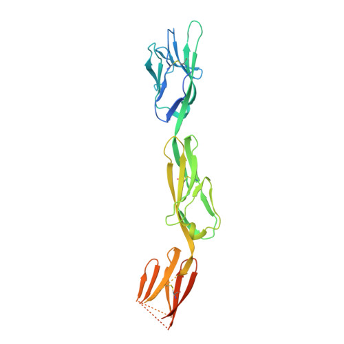 | |
UniProt & NIH Common Fund Data Resources | |||||
PHAROS: Q9P121 GTEx: ENSG00000182667 | |||||
Entity Groups | |||||
| Sequence Clusters | 30% Identity50% Identity70% Identity90% Identity95% Identity100% Identity | ||||
| UniProt Group | Q9P121 | ||||
Glycosylation | |||||
| Glycosylation Sites: 2 | Go to GlyGen: Q9P121-1 | ||||
Sequence AnnotationsExpand | |||||
Reference Sequence | |||||
| Ligands 1 Unique | |||||
|---|---|---|---|---|---|
| ID | Chains | Name / Formula / InChI Key | 2D Diagram | 3D Interactions | |
| NAG Download:Ideal Coordinates CCD File | D [auth A], E [auth A], F [auth A], G [auth B] | 2-acetamido-2-deoxy-beta-D-glucopyranose C8 H15 N O6 OVRNDRQMDRJTHS-FMDGEEDCSA-N |  | ||
| Length ( Å ) | Angle ( ˚ ) |
|---|---|
| a = 76.517 | α = 90 |
| b = 101.996 | β = 90 |
| c = 134.025 | γ = 90 |
| Software Name | Purpose |
|---|---|
| PHENIX | refinement |
| XDS | data reduction |
| Aimless | data scaling |
| PHASER | phasing |
| Funding Organization | Location | Grant Number |
|---|---|---|
| National Science Foundation (NSF, United States) | United States | MCB-1450895 |


