6DIK | pdb_00006dik

Crystal structure of Bothropstoxin I (BthTX-I) complexed to Chicoric acid


Experimental Data Snapshot

  • Method: X-RAY DIFFRACTION
  • Resolution: 1.93 Å
  • R-Value Free: 
    0.200 (Depositor), 0.200 (DCC) 
  • R-Value Work: 
    0.165 (Depositor), 0.166 (DCC) 
  • R-Value Observed: 
    0.166 (Depositor) 

Starting Model: experimental
View more details

wwPDB Validation 3D Report Full Report

Validation slider image for 6DIK

Ligand Structure Quality Assessment 


This is version 1.4 of the entry. See complete history

Literature

Structural basis of phospholipase A2-like myotoxin inhibition by chicoric acid, a novel potent inhibitor of ophidian toxins.

Cardoso, F.F.Borges, R.J.Dreyer, T.R.Salvador, G.H.M.Cavalcante, W.L.G.Pai, M.D.Gallacci, M.Fontes, M.R.M.

(2018) Biochim Biophys Acta Gen Subj 1862: 2728-2737

  • DOI: https://doi.org/10.1016/j.bbagen.2018.08.002
  • Primary Citation Related Structures: 
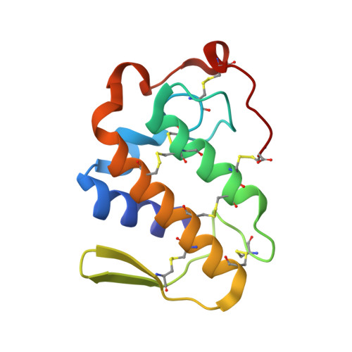
    6DIK

  • PubMed Abstract: 

    Specific compounds found in vegetal species have been demonstrated to be efficient inhibitors of snake toxins, such as phospholipase A 2 -like (PLA 2 -like) proteins. These particular proteins, present in several species of vipers (Viperidae), induce a severe local myotoxic effect in prey and human victims, and this effect is often not efficiently neutralized by the regular serum therapy. PLA 2 -like proteins have been functionally and structurally studied since the early 1990s; however, a comprehensive molecular mechanism was proposed only recently. Myographic and histological techniques were used to evaluate the inhibitory effect of chicoric acid (CA) against BthTX-I myotoxin. Isothermal titration calorimetry assays were used to measure the affinity between the inhibitor and the toxin. X-ray crystallography was used to reveal details of this interaction. CA prevented the blockade of indirectly evoked muscle contraction and inhibited muscle damage induced by BthTX-I. The inhibitor binds to the toxin with the highest affinity measured for a natural compound in calorimetric assays. The crystal structure and molecular dynamics simulations demonstrated that CA binds at the entrance of the hydrophobic channel of the toxin and binds to one of the clusters that participates in membrane disruption. CA prevents the myotoxic activity of the toxin, preventing its activation by simultaneous binding with two critical regions. CA is a potential myotoxic inhibitor to other PLA 2 -like proteins and a possible candidate to complement serum therapy.


  • Organizational Affiliation
    • Departamento de Física e Biofísica, Instituto de Biociências, Universidade Estadual Paulista (UNESP), Botucatu, SP, Brazil.

Macromolecule Content 

  • Total Structure Weight: 28.57 kDa 
  • Atom Count: 2,151 
  • Modeled Residue Count: 242 
  • Deposited Residue Count: 242 
  • Unique protein chains: 1

Macromolecules

Find similar proteins by:|  3D Structure
Entity ID: 1
MoleculeChains  Sequence LengthOrganismDetailsImage
Basic phospholipase A2 homolog bothropstoxin-1
A, B
121Bothrops jararacussuMutation(s): 0 
UniProt
Find proteins for Q90249 (Bothrops jararacussu)
Explore Q90249 
Go to UniProtKB:  Q90249
Entity Groups
Sequence Clusters30% Identity50% Identity70% Identity90% Identity95% Identity100% Identity
UniProt GroupQ90249
Sequence Annotations
Expand
Reference Sequence

Small Molecules

Ligands 4 Unique
IDChains Name / Formula / InChI Key2D Diagram3D Interactions
GKP

Query on GKP


Download:Ideal Coordinates CCD File
C [auth A](2R,3R)-2,3-bis{[(2E)-3-(3,4-dihydroxyphenyl)prop-2-enoyl]oxy}butanedioic acid
C22 H18 O12
YDDGKXBLOXEEMN-IABMMNSOSA-N
SO4

Query on SO4


Download:Ideal Coordinates CCD File
D [auth A],
E [auth A],
F [auth A],
G [auth B],
H [auth B]
SULFATE ION
O4 S
QAOWNCQODCNURD-UHFFFAOYSA-L
BCT

Query on BCT


Download:Ideal Coordinates CCD File
I [auth B]BICARBONATE ION
C H O3
BVKZGUZCCUSVTD-UHFFFAOYSA-M
EOH

Query on EOH


Download:Ideal Coordinates CCD File
J [auth B]ETHANOL
C2 H6 O
LFQSCWFLJHTTHZ-UHFFFAOYSA-N

Experimental Data & Validation

Experimental Data

  • Method: X-RAY DIFFRACTION
  • Resolution: 1.93 Å
  • R-Value Free:  0.200 (Depositor), 0.200 (DCC) 
  • R-Value Work:  0.165 (Depositor), 0.166 (DCC) 
  • R-Value Observed: 0.166 (Depositor) 
Space Group: P 1 21 1
Unit Cell:
Length ( Å )Angle ( ˚ )
a = 39.558α = 90
b = 71.632β = 109.02
c = 44.791γ = 90
Software Package:
Software NamePurpose
HKL-2000data reduction
SCALEPACKdata scaling
PHASERphasing
PHENIXrefinement
PDB_EXTRACTdata extraction

Structure Validation

View Full Validation Report


Ligand Structure Quality Assessment 


Entry History 

& Funding Information

Deposition Data


Funding OrganizationLocationGrant Number
Sao Paulo Research Foundation (FAPESP)Brazil2012/07112-6

Revision History  (Full details and data files)

  • Version 1.0: 2018-10-03
    Type: Initial release
  • Version 1.1: 2018-10-31
    Changes: Data collection, Database references
  • Version 1.2: 2020-01-01
    Changes: Author supporting evidence
  • Version 1.3: 2023-10-11
    Changes: Advisory, Data collection, Database references, Refinement description
  • Version 1.4: 2024-11-06
    Changes: Structure summary