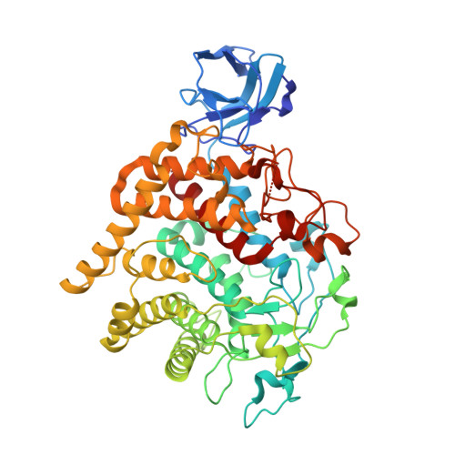
6DHT

Bacteroides ovatus GH9 Bacova_02649


Experimental Data Snapshot

  • Method: X-RAY DIFFRACTION
  • Resolution: 1.42 Å
  • R-Value Free: 0.197 
  • R-Value Work: 0.168 
  • R-Value Observed: 0.168 

wwPDB Validation   3D Report Full Report


This is version 1.2 of the entry. See complete history


Literature

A Cell-Surface GH9 Endo-Glucanase Coordinates with Surface Glycan-Binding Proteins to Mediate Xyloglucan Uptake in the Gut Symbiont Bacteroides ovatus.

Foley, M.H.Dejean, G.Hemsworth, G.R.Davies, G.J.Brumer, H.Koropatkin, N.M.

(2019) J Mol Biol 431: 981-995

  • DOI: https://doi.org/10.1016/j.jmb.2019.01.008
  • Primary Citation of Related Structures:  
    6DHT

  • PubMed Abstract: 

    Dietary fiber is an important food source for members of the human gut microbiome. Members of the dominant Bacteroidetes phylum capture diverse polysaccharides via the action of multiple cell surface proteins encoded within polysaccharide utilization loci (PUL). The independent activities of PUL-encoded glycoside hydrolases (GHs) and surface glycan-binding proteins (SGBPs) for the harvest of various glycans have been studied in detail, but how these proteins work together to coordinate uptake is poorly understood. Here, we combine genetic and biochemical approaches to discern the interplay between the BoGH9 endoglucanase and the xyloglucan-binding proteins SGBP-A and SGBP-B from the Bacteroides ovatus xyloglucan utilization locus (XyGUL). The expression of BoGH9, a weakly active xyloglucanase in isolation, is required in a strain that expresses a non-binding version of SGBP-A (SGBP-A*). The crystal structure of the BoGH9 enzyme suggests the molecular basis for its robust activity on mixed-linkage β-glucan compared to xyloglucan. However, catalytically inactive site-directed mutants of BoGH9 fail to complement the deletion of the active BoGH9 in a SGBP-A* strain. We also find that SGBP-B is needed in an SGBP-A* background to support growth on xyloglucan, but that the non-binding SGBP-B* protein acts in a dominant negative manner to inhibit growth on xyloglucan. We postulate a model whereby the SGBP-A, SGBP-B, and BoGH9 work together at the cell surface, likely within a discrete complex, and that xyloglucan binding by SGBP-B and BoGH9 may facilitate the orientation of the xyloglucan for transfer across the outer membrane.


  • Organizational Affiliation

    Department of Microbiology and Immunology, University of Michigan Medical School, Ann Arbor, MI 48109, USA.


Macromolecules
Find similar proteins by:  (by identity cutoff)  |  3D Structure
Entity ID: 1
MoleculeChains Sequence LengthOrganismDetailsImage
Xyloglucan-specific endo-beta-1,4-glucanase BoGH9A569Bacteroides ovatus ATCC 8483Mutation(s): 0 
Gene Names: BACOVA_02649
EC: 3.2.1.151
UniProt
Find proteins for A7LXT3 (Bacteroides ovatus (strain ATCC 8483 / DSM 1896 / JCM 5824 / BCRC 10623 / CCUG 4943 / NCTC 11153))
Explore A7LXT3 
Go to UniProtKB:  A7LXT3
Entity Groups  
Sequence Clusters30% Identity50% Identity70% Identity90% Identity95% Identity100% Identity
UniProt GroupA7LXT3
Sequence Annotations
Expand
  • Reference Sequence
Small Molecules
Ligands 6 Unique
IDChains Name / Formula / InChI Key2D Diagram3D Interactions
PEG
Query on PEG

Download Ideal Coordinates CCD File 
S [auth A],
T [auth A],
U [auth A]
DI(HYDROXYETHYL)ETHER
C4 H10 O3
MTHSVFCYNBDYFN-UHFFFAOYSA-N
IMD
Query on IMD

Download Ideal Coordinates CCD File 
B [auth A]IMIDAZOLE
C3 H5 N2
RAXXELZNTBOGNW-UHFFFAOYSA-O
EDO
Query on EDO

Download Ideal Coordinates CCD File 
E [auth A]
F [auth A]
G [auth A]
H [auth A]
I [auth A]
E [auth A],
F [auth A],
G [auth A],
H [auth A],
I [auth A],
J [auth A],
K [auth A],
L [auth A],
M [auth A],
N [auth A],
O [auth A],
P [auth A],
Q [auth A],
R [auth A]
1,2-ETHANEDIOL
C2 H6 O2
LYCAIKOWRPUZTN-UHFFFAOYSA-N
CA
Query on CA

Download Ideal Coordinates CCD File 
C [auth A],
D [auth A]
CALCIUM ION
Ca
BHPQYMZQTOCNFJ-UHFFFAOYSA-N
CL
Query on CL

Download Ideal Coordinates CCD File 
V [auth A],
W [auth A]
CHLORIDE ION
Cl
VEXZGXHMUGYJMC-UHFFFAOYSA-M
MG
Query on MG

Download Ideal Coordinates CCD File 
X [auth A]MAGNESIUM ION
Mg
JLVVSXFLKOJNIY-UHFFFAOYSA-N
Experimental Data & Validation

Experimental Data

  • Method: X-RAY DIFFRACTION
  • Resolution: 1.42 Å
  • R-Value Free: 0.197 
  • R-Value Work: 0.168 
  • R-Value Observed: 0.168 
  • Space Group: C 1 2 1
Unit Cell:
Length ( Å )Angle ( ˚ )
a = 130.646α = 90
b = 50.406β = 109
c = 95.98γ = 90
Software Package:
Software NamePurpose
HKL-2000data reduction
SCALEPACKdata scaling
PHASERphasing
PHENIXrefinement
PDB_EXTRACTdata extraction

Structure Validation

View Full Validation Report


Entry History & Funding Information

Deposition Data


Funding OrganizationLocationGrant Number
National Institutes of Health/National Institute of General Medical Sciences (NIH/NIGMS)United StatesGM118475

Revision History  (Full details and data files)

  • Version 1.0: 2019-04-03
    Type: Initial release
  • Version 1.1: 2020-01-01
    Changes: Author supporting evidence
  • Version 1.2: 2024-03-13
    Changes: Data collection, Database references, Derived calculations