6DC2 | pdb_00006dc2

Crystal structure of Desulfovibrio vulgaris carbon monoxide dehydrogenase C301S variant


Experimental Data Snapshot

  • Method: X-RAY DIFFRACTION
  • Resolution: 1.99 Å
  • R-Value Free: 
    0.212 (Depositor), 0.213 (DCC) 
  • R-Value Work: 
    0.163 (Depositor), 0.165 (DCC) 
  • R-Value Observed: 
    0.166 (Depositor) 

Starting Model: experimental
View more details

wwPDB Validation 3D Report Full Report

Validation slider image for 6DC2

This is version 1.3 of the entry. See complete history

Literature

Redox-dependent rearrangements of the NiFeS cluster of carbon monoxide dehydrogenase.

Wittenborn, E.C.Merrouch, M.Ueda, C.Fradale, L.Leger, C.Fourmond, V.Pandelia, M.E.Dementin, S.Drennan, C.L.

(2018) Elife 7

  • DOI: https://doi.org/10.7554/eLife.39451
  • Primary Citation Related Structures: 
    6B6V, 6B6W, 6B6X, 6B6Y, 6DC2

  • PubMed Abstract: 

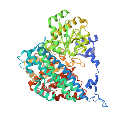
    The C-cluster of the enzyme carbon monoxide dehydrogenase (CODH) is a structurally distinctive Ni-Fe-S cluster employed to catalyze the reduction of CO 2 to CO as part of the Wood-Ljungdahl carbon fixation pathway. Using X-ray crystallography, we have observed unprecedented conformational dynamics in the C-cluster of the CODH from Desulfovibrio vulgaris , providing the first view of an oxidized state of the cluster. Combined with supporting spectroscopic data, our structures reveal that this novel, oxidized cluster arrangement plays a role in avoiding irreversible oxidative degradation at the C-cluster. Furthermore, mutagenesis of a conserved cysteine residue that binds the C-cluster in the oxidized state but not in the reduced state suggests that the oxidized conformation could be important for proper cluster assembly, in particular Ni incorporation. Together, these results lay a foundation for future investigations of C-cluster activation and assembly, and contribute to an emerging paradigm of metallocluster plasticity.


  • Organizational Affiliation
    • Department of Chemistry, Massachusetts Institute of Technology, Cambridge, United States.

Macromolecule Content 

  • Total Structure Weight: 274.91 kDa 
  • Atom Count: 20,110 
  • Modeled Residue Count: 2,499 
  • Deposited Residue Count: 2,548 
  • Unique protein chains: 1

Macromolecules

Find similar proteins by:|  3D Structure
Entity ID: 1
MoleculeChains  Sequence LengthOrganismDetailsImage
Carbon monoxide dehydrogenase
A, B, C, D
637Nitratidesulfovibrio vulgarisMutation(s): 1 
Gene Names: cooSDVU_2098
EC: 1.2.7.4
UniProt
Find proteins for Q72A99 (Nitratidesulfovibrio vulgaris (strain ATCC 29579 / DSM 644 / CCUG 34227 / NCIMB 8303 / VKM B-1760 / Hildenborough))
Explore Q72A99 
Go to UniProtKB:  Q72A99
Entity Groups
Sequence Clusters30% Identity50% Identity70% Identity90% Identity95% Identity100% Identity
UniProt GroupQ72A99
Sequence Annotations
Expand
Reference Sequence

Small Molecules

Ligands 6 Unique
IDChains Name / Formula / InChI Key2D Diagram3D Interactions
XCC
(Subject of Investigation/LOI)

Query on XCC


Download:Ideal Coordinates CCD File
G [auth A],
L [auth B],
R [auth C],
Y [auth D]
FE(4)-NI(1)-S(4) CLUSTER
Fe4 Ni S4
QGLWBXDZIHZONR-UHFFFAOYSA-N
SF4

Query on SF4


Download:Ideal Coordinates CCD File
E [auth A],
K [auth B],
P [auth C],
X [auth D]
IRON/SULFUR CLUSTER
Fe4 S4
LJBDFODJNLIPKO-UHFFFAOYSA-N
FES

Query on FES


Download:Ideal Coordinates CCD File
F [auth A],
Q [auth C]
FE2/S2 (INORGANIC) CLUSTER
Fe2 S2
NIXDOXVAJZFRNF-UHFFFAOYSA-N
GOL

Query on GOL


Download:Ideal Coordinates CCD File
N [auth B],
T [auth C],
U [auth C],
V [auth C]
GLYCEROL
C3 H8 O3
PEDCQBHIVMGVHV-UHFFFAOYSA-N
FE

Query on FE


Download:Ideal Coordinates CCD File
H [auth A],
M [auth B],
S [auth C],
Z [auth D]
FE (III) ION
Fe
VTLYFUHAOXGGBS-UHFFFAOYSA-N
MG

Query on MG


Download:Ideal Coordinates CCD File
AA [auth D],
I [auth A],
J [auth A],
O [auth B],
W [auth C]
MAGNESIUM ION
Mg
JLVVSXFLKOJNIY-UHFFFAOYSA-N

Experimental Data & Validation

Experimental Data

  • Method: X-RAY DIFFRACTION
  • Resolution: 1.99 Å
  • R-Value Free:  0.212 (Depositor), 0.213 (DCC) 
  • R-Value Work:  0.163 (Depositor), 0.165 (DCC) 
  • R-Value Observed: 0.166 (Depositor) 
Space Group: P 1 21 1
Unit Cell:
Length ( Å )Angle ( ˚ )
a = 64.79α = 90
b = 144.15β = 98.5
c = 123.69γ = 90
Software Package:
Software NamePurpose
PHENIXrefinement
XDSdata reduction
XSCALEdata scaling
PHASERphasing

Structure Validation

View Full Validation Report


Entry History 

& Funding Information

Deposition Data


Funding OrganizationLocationGrant Number
National Institutes of Health/National Institute of General Medical Sciences (NIH/NIGMS)United StatesGM069857
National Institutes of Health/National Institute of General Medical Sciences (NIH/NIGMS)United StatesT32 GM008334

Revision History  (Full details and data files)

  • Version 1.0: 2018-10-03
    Type: Initial release
  • Version 1.1: 2018-10-17
    Changes: Data collection, Database references
  • Version 1.2: 2020-01-01
    Changes: Author supporting evidence
  • Version 1.3: 2023-10-04
    Changes: Data collection, Database references, Derived calculations, Refinement description