


Crystal structure of PT1940 bound to HIF2a-B*:ARNT-B* complex
Du, X.To be published.
Experimental Data Snapshot
Starting Model: experimental
View more details
Entity ID: 1 | |||||
|---|---|---|---|---|---|
| Molecule | Chains | Sequence Length | Organism | Details | Image |
| Endothelial PAS domain-containing protein 1 | 117 | Homo sapiens | Mutation(s): 1 Gene Names: EPAS1, BHLHE73, HIF2A, MOP2, PASD2 | 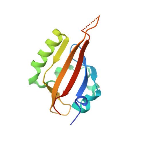 | |
UniProt & NIH Common Fund Data Resources | |||||
PHAROS: Q99814 GTEx: ENSG00000116016 | |||||
Entity Groups | |||||
| Sequence Clusters | 30% Identity50% Identity70% Identity90% Identity95% Identity100% Identity | ||||
| UniProt Group | Q99814 | ||||
Sequence AnnotationsExpand | |||||
Reference Sequence | |||||
Entity ID: 2 | |||||
|---|---|---|---|---|---|
| Molecule | Chains | Sequence Length | Organism | Details | Image |
| Aryl hydrocarbon receptor nuclear translocator | 121 | Homo sapiens | Mutation(s): 1 Gene Names: ARNT, BHLHE2 | 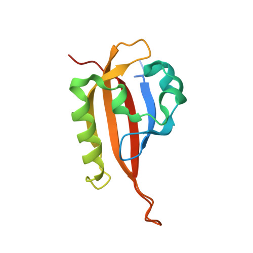 | |
UniProt & NIH Common Fund Data Resources | |||||
PHAROS: P27540 GTEx: ENSG00000143437 | |||||
Entity Groups | |||||
| Sequence Clusters | 30% Identity50% Identity70% Identity90% Identity95% Identity100% Identity | ||||
| UniProt Group | P27540 | ||||
Sequence AnnotationsExpand | |||||
Reference Sequence | |||||
| Ligands 1 Unique | |||||
|---|---|---|---|---|---|
| ID | Chains | Name / Formula / InChI Key | 2D Diagram | 3D Interactions | |
| FOJ Download:Ideal Coordinates CCD File | C [auth A] | 3-{[(3R)-4-(difluoromethyl)-2,2-difluoro-3-hydroxy-1,1-dioxo-2,3-dihydro-1H-1-benzothiophen-5-yl]oxy}-5-fluorobenzonitrile C16 H8 F5 N O4 S HZDKYXAZAPXCKQ-CQSZACIVSA-N |  | ||
| Length ( Å ) | Angle ( ˚ ) |
|---|---|
| a = 73.216 | α = 90 |
| b = 83.832 | β = 106.25 |
| c = 41.41 | γ = 90 |
| Software Name | Purpose |
|---|---|
| REFMAC | refinement |
| HKL-3000 | data reduction |
| SCALEPACK | data scaling |
| PDB_EXTRACT | data extraction |
| PHASER | phasing |


