6CVM

Atomic resolution cryo-EM structure of beta-galactosidase


Experimental Data Snapshot

  • Method: ELECTRON MICROSCOPY
  • Resolution: 1.90 Å
  • Aggregation State: PARTICLE 
  • Reconstruction Method: SINGLE PARTICLE 

wwPDB Validation   3D Report Full Report


This is version 1.4 of the entry. See complete history


Literature

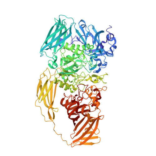
Atomic Resolution Cryo-EM Structure of beta-Galactosidase.

Bartesaghi, A.Aguerrebere, C.Falconieri, V.Banerjee, S.Earl, L.A.Zhu, X.Grigorieff, N.Milne, J.L.S.Sapiro, G.Wu, X.Subramaniam, S.

(2018) Structure 26: 848

  • DOI: https://doi.org/10.1016/j.str.2018.04.004
  • Primary Citation of Related Structures:  
    6CVM

  • PubMed Abstract: 

    The advent of direct electron detectors has enabled the routine use of single-particle cryo-electron microscopy (EM) approaches to determine structures of a variety of protein complexes at near-atomic resolution. Here, we report the development of methods to account for local variations in defocus and beam-induced drift, and the implementation of a data-driven dose compensation scheme that significantly improves the extraction of high-resolution information recorded during exposure of the specimen to the electron beam. These advances enable determination of a cryo-EM density map for β-galactosidase bound to the inhibitor phenylethyl β-D-thiogalactopyranoside where the ordered regions are resolved at a level of detail seen in X-ray maps at ∼ 1.5 Å resolution. Using this density map in conjunction with constrained molecular dynamics simulations provides a measure of the local flexibility of the non-covalently bound inhibitor and offers further opportunities for structure-guided inhibitor design.


  • Organizational Affiliation

    Laboratory of Cell Biology, Center for Cancer Research, National Cancer Institute, NIH, Bethesda, MD 20892, USA.


Macromolecules
Find similar proteins by:  (by identity cutoff)  |  3D Structure
Entity ID: 1
MoleculeChains Sequence LengthOrganismDetailsImage
Beta-galactosidase
A, B, C, D
1,021Escherichia coli K-12Mutation(s): 0 
EC: 3.2.1.23
UniProt
Find proteins for P00722 (Escherichia coli (strain K12))
Explore P00722 
Go to UniProtKB:  P00722
Entity Groups  
Sequence Clusters30% Identity50% Identity70% Identity90% Identity95% Identity100% Identity
UniProt GroupP00722
Sequence Annotations
Expand
  • Reference Sequence
Small Molecules
Ligands 3 Unique
IDChains Name / Formula / InChI Key2D Diagram3D Interactions
PTQ
Query on PTQ

Download Ideal Coordinates CCD File 
E [auth A],
J [auth B],
O [auth C],
T [auth D]
2-phenylethyl 1-thio-beta-D-galactopyranoside
C14 H20 O5 S
ZNAMMSOYKPMPGC-HTOAHKCRSA-N
MG
Query on MG

Download Ideal Coordinates CCD File 
F [auth A]
G [auth A]
K [auth B]
L [auth B]
P [auth C]
F [auth A],
G [auth A],
K [auth B],
L [auth B],
P [auth C],
Q [auth C],
U [auth D],
V [auth D]
MAGNESIUM ION
Mg
JLVVSXFLKOJNIY-UHFFFAOYSA-N
NA
Query on NA

Download Ideal Coordinates CCD File 
H [auth A]
I [auth A]
M [auth B]
N [auth B]
R [auth C]
H [auth A],
I [auth A],
M [auth B],
N [auth B],
R [auth C],
S [auth C],
W [auth D],
X [auth D]
SODIUM ION
Na
FKNQFGJONOIPTF-UHFFFAOYSA-N
Experimental Data & Validation

Experimental Data

  • Method: ELECTRON MICROSCOPY
  • Resolution: 1.90 Å
  • Aggregation State: PARTICLE 
  • Reconstruction Method: SINGLE PARTICLE 
EM Software:
TaskSoftware PackageVersion
MODEL REFINEMENTPHENIX
RECONSTRUCTIONFREALIGN9.11

Structure Validation

View Full Validation Report


Entry History 

Deposition Data

Revision History  (Full details and data files)

  • Version 1.0: 2018-05-30
    Type: Initial release
  • Version 1.1: 2018-06-20
    Changes: Data collection, Database references
  • Version 1.2: 2018-07-18
    Changes: Data collection
  • Version 1.3: 2018-08-22
    Changes: Data collection, Database references
  • Version 1.4: 2024-03-13
    Changes: Data collection, Database references, Derived calculations