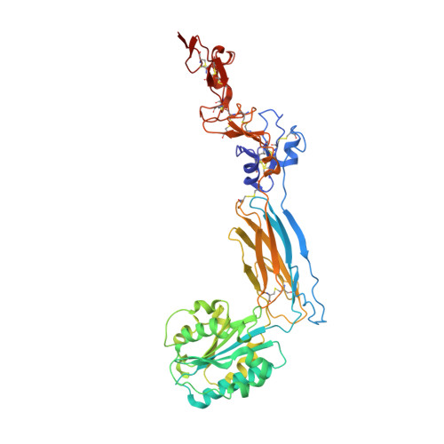
6CKB

Crystal structure of an extended beta3 integrin P33

  • Classification: CELL ADHESION
  • Organism(s): Homo sapiens
  • Expression System: Homo sapiens
  • Mutation(s): No 

  • Deposited: 2018-02-27 Released: 2018-08-01 
  • Deposition Author(s): Zhou, D., Zhu, J.
  • Funding Organization(s): National Institutes of Health/National Heart, Lung, and Blood Institute (NIH/NHLBI)

Experimental Data Snapshot

  • Method: X-RAY DIFFRACTION
  • Resolution: 2.80 Å
  • R-Value Free: 0.296 
  • R-Value Work: 0.239 
  • R-Value Observed: 0.242 

wwPDB Validation   3D Report Full Report


Ligand Structure Quality Assessment 


This is version 1.4 of the entry. See complete history


Literature

Structure of an extended beta3integrin.

Zhou, D.Thinn, A.M.M.Zhao, Y.Wang, Z.Zhu, J.

(2018) Blood 132: 962-972

  • DOI: https://doi.org/10.1182/blood-2018-01-829572
  • Primary Citation of Related Structures:  
    6BXB, 6BXF, 6CKB

  • PubMed Abstract: 

    Cells use adhesion receptor integrins to communicate with their surroundings. Integrin activation and cellular signaling are coupled with change from bent to extended conformation. β 3 integrins, including α IIb β 3 , which is essential for the function of platelets in hemostasis and thrombosis, and α V β 3 , which plays multiple roles in diverse cell types, have been prototypes in understanding integrin structure and function. Despite extensive structural studies, a high-resolution integrin structure in an extended conformation remains to be determined. The human β 3 Leu33Pro polymorphism, located at the PSI domain, defines human platelet-specific alloantigens 1a and 1b (HPA-1a/b), immune response to which is a cause of posttransfusion purpura and fetal/neonatal alloimmune thrombocytopenia. Leu33Pro substitution has also been suggested to be a risk factor for thrombosis. Here we report the crystal structure of the β 3 headpiece in either Leu33 or Pro33 form, both of which reveal intermediate and fully extended conformations coexisting in 1 crystal. These were used to build high-resolution structures of full-length β 3 integrin in the intermediate and fully extended states, agreeing well with the corresponding conformations observed by electron microscopy. Our structures reveal how β 3 integrin becomes extended at its β-knee region and how the flexibility of β-leg domains is determined. In addition, our structures reveal conformational changes of the PSI and I-EGF1 domains upon β 3 extension, which may affect the binding of conformation-dependent anti-HPA-1a alloantibodies. Our structural and functional data show that Leu33Pro substitution does not directly alter the conformation or ligand binding of β 3 integrin.


  • Organizational Affiliation

    Blood Research Institute, BloodCenter of Wisconsin, part of Versiti, Milwaukee, WI.


Macromolecules
Find similar proteins by:  (by identity cutoff)  |  3D Structure
Entity ID: 1
MoleculeChains Sequence LengthOrganismDetailsImage
Chimera protein of Integrin beta-3 and Integrin alpha-L
A, B
466Homo sapiensMutation(s): 0 
Gene Names: ITGB3GP3AITGALCD11A
UniProt & NIH Common Fund Data Resources
Find proteins for P20701 (Homo sapiens)
Explore P20701 
Go to UniProtKB:  P20701
PHAROS:  P20701
GTEx:  ENSG00000005844 
Find proteins for P05106 (Homo sapiens)
Explore P05106 
Go to UniProtKB:  P05106
PHAROS:  P05106
GTEx:  ENSG00000259207 
Entity Groups  
Sequence Clusters30% Identity50% Identity70% Identity90% Identity95% Identity100% Identity
UniProt GroupsP20701P05106
Glycosylation
Glycosylation Sites: 2Go to GlyGen: P05106-1P20701-1
Sequence Annotations
Expand
  • Reference Sequence
Small Molecules
Ligands 3 Unique
IDChains Name / Formula / InChI Key2D Diagram3D Interactions
NAG
Query on NAG

Download Ideal Coordinates CCD File 
D [auth A],
E [auth A],
F [auth B],
G [auth B],
H [auth B]
2-acetamido-2-deoxy-beta-D-glucopyranose
C8 H15 N O6
OVRNDRQMDRJTHS-FMDGEEDCSA-N
CA
Query on CA

Download Ideal Coordinates CCD File 
I [auth B]CALCIUM ION
Ca
BHPQYMZQTOCNFJ-UHFFFAOYSA-N
MG
Query on MG

Download Ideal Coordinates CCD File 
C [auth A]MAGNESIUM ION
Mg
JLVVSXFLKOJNIY-UHFFFAOYSA-N
Experimental Data & Validation

Experimental Data

  • Method: X-RAY DIFFRACTION
  • Resolution: 2.80 Å
  • R-Value Free: 0.296 
  • R-Value Work: 0.239 
  • R-Value Observed: 0.242 
  • Space Group: P 1 21 1
Unit Cell:
Length ( Å )Angle ( ˚ )
a = 59.08α = 90
b = 80.91β = 96.36
c = 126.72γ = 90
Software Package:
Software NamePurpose
PHENIXrefinement
MOSFLMdata reduction
SCALAdata scaling
PHASERphasing

Structure Validation

View Full Validation Report


Ligand Structure Quality Assessment 


Entry History & Funding Information

Deposition Data


Funding OrganizationLocationGrant Number
National Institutes of Health/National Heart, Lung, and Blood Institute (NIH/NHLBI)United StatesR01HL131386

Revision History  (Full details and data files)

  • Version 1.0: 2018-08-01
    Type: Initial release
  • Version 1.1: 2018-09-12
    Changes: Data collection, Database references
  • Version 1.2: 2019-12-04
    Changes: Author supporting evidence
  • Version 1.3: 2020-07-29
    Type: Remediation
    Reason: Carbohydrate remediation
    Changes: Data collection, Derived calculations, Structure summary
  • Version 1.4: 2024-10-23
    Changes: Data collection, Database references, Structure summary