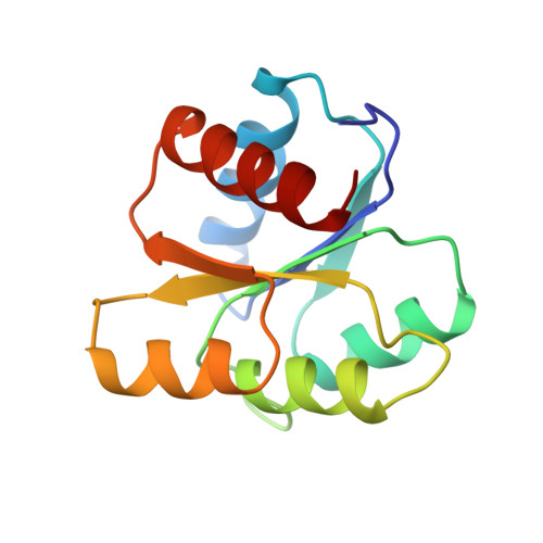
6CHY

STRUCTURE OF CHEMOTAXIS PROTEIN CHEY


Experimental Data Snapshot

  • Method: X-RAY DIFFRACTION
  • Resolution: 2.33 Å
  • R-Value Work: 0.185 

Starting Model: experimental
View more details

wwPDB Validation   3D Report Full Report


This is version 1.4 of the entry. See complete history


Literature

Crystal structures of CheY mutants Y106W and T87I/Y106W. CheY activation correlates with movement of residue 106.

Zhu, X.Rebello, J.Matsumura, P.Volz, K.

(1997) J Biol Chem 272: 5000-5006

  • DOI: https://doi.org/10.1074/jbc.272.8.5000
  • Primary Citation of Related Structures:  
    5CHY, 6CHY

  • PubMed Abstract: 

    Position 106 in CheY is highly conserved as an aromatic residue in the response regulator superfamily. In the structure of the wild-type, apo-CheY, Tyr106 is a rotamer whose electron density is observed in both the inside and the outside positions. In the structure of the T87I mutant of CheY, the threonine to isoleucine change at position 87 causes the side chain of Tyr106 to be exclusively restricted to the outside position. In this report we demonstrate that the T87I mutation causes cells to be smooth swimming and non-chemotactic. We also show that another CheY mutant, Y106W, causes cells to be more tumbly than wild-type CheY, and impairs chemotaxis. In the structure of Y106W, the side chain of Trp106 stays exclusively in the inside position. Furthermore, a T87I/Y106W double mutant, which confers the same phenotype as T87I, restricts the side chain of Trp106 to the outside position. The results from these behavioral and structural studies indicate that the rotameric nature of the Tyr106 residue is involved in activation of the CheY molecule. Specifically, CheY's signaling ability correlates with the conformational heterogeneity of the Tyr106 side chain. Our data also suggest that these mutations affect the signal at an event subsequent to phosphorylation.


  • Organizational Affiliation

    Department of Microbiology and Immunology, University of Illinois, Chicago, Illinois 60612, USA.


Macromolecules
Find similar proteins by:  (by identity cutoff)  |  3D Structure
Entity ID: 1
MoleculeChains Sequence LengthOrganismDetailsImage
CHEY
A, B
128Escherichia coli K-12Mutation(s): 2 
UniProt
Find proteins for P0AE67 (Escherichia coli (strain K12))
Explore P0AE67 
Go to UniProtKB:  P0AE67
Entity Groups  
Sequence Clusters30% Identity50% Identity70% Identity90% Identity95% Identity100% Identity
UniProt GroupP0AE67
Sequence Annotations
Expand
  • Reference Sequence
Small Molecules
Ligands 1 Unique
IDChains Name / Formula / InChI Key2D Diagram3D Interactions
SO4
Query on SO4

Download Ideal Coordinates CCD File 
C [auth B]SULFATE ION
O4 S
QAOWNCQODCNURD-UHFFFAOYSA-L
Experimental Data & Validation

Experimental Data

  • Method: X-RAY DIFFRACTION
  • Resolution: 2.33 Å
  • R-Value Work: 0.185 
  • Space Group: P 21 21 2
Unit Cell:
Length ( Å )Angle ( ˚ )
a = 95.78α = 90
b = 76.7β = 90
c = 32.64γ = 90
Software Package:
Software NamePurpose
MERLOTphasing
PROFFTrefinement
MADNESdata reduction
MADNESdata scaling

Structure Validation

View Full Validation Report


Entry History 

Deposition Data

Revision History  (Full details and data files)

  • Version 1.0: 1996-12-07
    Type: Initial release
  • Version 1.1: 2008-03-25
    Changes: Version format compliance
  • Version 1.2: 2011-07-13
    Changes: Version format compliance
  • Version 1.3: 2021-11-03
    Changes: Database references, Derived calculations
  • Version 1.4: 2024-05-22
    Changes: Data collection, Refinement description