6CEF

Crystal structure of fragment 3-(1,3-Benzothiazol-2-yl)propanoic acid bound in the ubiquitin binding pocket of the HDAC6 zinc-finger domain


Experimental Data Snapshot

  • Method: X-RAY DIFFRACTION
  • Resolution: 1.80 Å
  • R-Value Free: 
    0.181 (Depositor), 0.190 (DCC) 
  • R-Value Work: 
    0.150 (Depositor), 0.160 (DCC) 
  • R-Value Observed: 
    0.151 (Depositor) 

Starting Model: experimental
View more details

wwPDB Validation   3D Report Full Report


Ligand Structure Quality Assessment 

Created with Raphaël 2.3.0Worse 01 BetterLigand structure goodness of fit to experimental dataBest fitted EYJClick on this verticalbar to view details

This is version 1.3 of the entry. See complete history


Literature

Identification and Structure-Activity Relationship of HDAC6 Zinc-Finger Ubiquitin Binding Domain Inhibitors.

Ferreira de Freitas, R.Harding, R.J.Franzoni, I.Ravichandran, M.Mann, M.K.Ouyang, H.Lautens, M.Santhakumar, V.Arrowsmith, C.H.Schapira, M.

(2018) J Med Chem 61: 4517-4527

  • DOI: https://doi.org/10.1021/acs.jmedchem.8b00258
  • Primary Citation of Related Structures:  
    6CE6, 6CE8, 6CEA, 6CEC, 6CED, 6CEE, 6CEF

  • PubMed Abstract: 

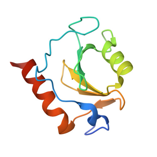
    HDAC6 plays a central role in the recruitment of protein aggregates for lysosomal degradation and is a promising target for combination therapy with proteasome inhibitors in multiple myeloma. Pharmacologically displacing ubiquitin from the zinc-finger ubiquitin-binding domain (ZnF-UBD) of HDAC6 is an underexplored alternative to catalytic inhibition. Here, we present the discovery of an HDAC6 ZnF-UBD-focused chemical series and its progression from virtual screening hits to low micromolar inhibitors. A carboxylate mimicking the C-terminal extremity of ubiquitin, and an extended aromatic system stacking with W1182 and R1155, are necessary for activity. One of the compounds induced a conformational remodeling of the binding site where the primary binding pocket opens up onto a ligand-able secondary pocket that may be exploited to increase potency. The preliminary structure-activity relationship accompanied by nine crystal structures should enable further optimization into a chemical probe to investigate the merit of targeting the ZnF-UBD of HDAC6 in multiple myeloma and other diseases.


  • Organizational Affiliation

    Structural Genomics Consortium , University of Toronto , MaRS South Tower, Suite 700, 101 College Street , Toronto , Ontario M5G 1L7 , Canada.


Macromolecules
Find similar proteins by:  (by identity cutoff)  |  3D Structure
Entity ID: 1
MoleculeChains Sequence LengthOrganismDetailsImage
Histone deacetylase 6107Homo sapiensMutation(s): 0 
Gene Names: HDAC6KIAA0901JM21
EC: 3.5.1.98 (PDB Primary Data), 3.5.1 (UniProt)
UniProt & NIH Common Fund Data Resources
Find proteins for Q9UBN7 (Homo sapiens)
Explore Q9UBN7 
Go to UniProtKB:  Q9UBN7
PHAROS:  Q9UBN7
GTEx:  ENSG00000094631 
Entity Groups  
Sequence Clusters30% Identity50% Identity70% Identity90% Identity95% Identity100% Identity
UniProt GroupQ9UBN7
Sequence Annotations
Expand
  • Reference Sequence
Small Molecules
Ligands 3 Unique
IDChains Name / Formula / InChI Key2D Diagram3D Interactions
EYJ
Query on EYJ

Download Ideal Coordinates CCD File 
H [auth A]3-(1,3-benzothiazol-2-yl)propanoic acid
C10 H9 N O2 S
WHNQTHDJEZTVHS-UHFFFAOYSA-N
ZN
Query on ZN

Download Ideal Coordinates CCD File 
B [auth A],
C [auth A],
D [auth A]
ZINC ION
Zn
PTFCDOFLOPIGGS-UHFFFAOYSA-N
UNX
Query on UNX

Download Ideal Coordinates CCD File 
E [auth A],
F [auth A],
G [auth A]
UNKNOWN ATOM OR ION
X
Binding Affinity Annotations 
IDSourceBinding Affinity
EYJ BindingDB:  6CEF IC50: 1.80e+5 (nM) from 1 assay(s)
Experimental Data & Validation

Experimental Data

  • Method: X-RAY DIFFRACTION
  • Resolution: 1.80 Å
  • R-Value Free:  0.181 (Depositor), 0.190 (DCC) 
  • R-Value Work:  0.150 (Depositor), 0.160 (DCC) 
  • R-Value Observed: 0.151 (Depositor) 
Space Group: P 21 21 21
Unit Cell:
Length ( Å )Angle ( ˚ )
a = 40.7α = 90
b = 44.21β = 90
c = 56.03γ = 90
Software Package:
Software NamePurpose
REFMACrefinement
Aimlessdata scaling
PDB_EXTRACTdata extraction
xia2data reduction

Structure Validation

View Full Validation Report


Ligand Structure Quality Assessment 

Created with Raphaël 2.3.0Worse 01 BetterLigand structure goodness of fit to experimental dataBest fitted EYJClick on this verticalbar to view details

Entry History 

Revision History  (Full details and data files)

  • Version 1.0: 2018-02-28
    Type: Initial release
  • Version 1.1: 2018-05-16
    Changes: Data collection, Database references
  • Version 1.2: 2018-05-30
    Changes: Data collection, Database references
  • Version 1.3: 2023-10-04
    Changes: Data collection, Database references, Refinement description