6CDM | pdb_00006cdm

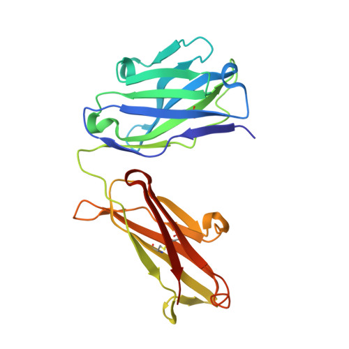
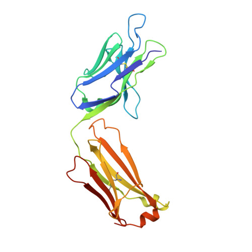
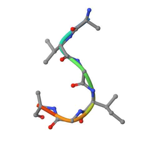
Structure of vaccine-elicited HIV-1 neutralizing antibody vFP7.04 in complex with HIV-1 fusion peptide residue 512-519


Experimental Data Snapshot

  • Method: X-RAY DIFFRACTION
  • Resolution: 2.41 Å
  • R-Value Free: 
    0.222 (Depositor), 0.223 (DCC) 
  • R-Value Work: 
    0.183 (Depositor), 0.184 (DCC) 
  • R-Value Observed: 
    0.185 (Depositor) 

Starting Model: experimental
View more details

wwPDB Validation 3D Report Full Report

Validation slider image for 6CDM

This is version 1.3 of the entry. See complete history. 

Literature

Epitope-based vaccine design yields fusion peptide-directed antibodies that neutralize diverse strains of HIV-1.

Xu, K., Acharya, P., Kong, R., Cheng, C., Chuang, G.Y., Liu, K., Louder, M.K., O'Dell, S., Rawi, R., Sastry, M., Shen, C.H., Zhang, B., Zhou, T., Asokan, M., Bailer, R.T., Chambers, M., Chen, X., Choi, C.W., Dandey, V.P., Doria-Rose, N.A., Druz, A., Eng, E.T., Farney, S.K., Foulds, K.E., Geng, H., Georgiev, I.S., Gorman, J., Hill, K.R., Jafari, A.J., Kwon, Y.D., Lai, Y.T., Lemmin, T., McKee, K., Ohr, T.Y., Ou, L., Peng, D., Rowshan, A.P., Sheng, Z., Todd, J.P., Tsybovsky, Y., Viox, E.G., Wang, Y., Wei, H., Yang, Y., Zhou, A.F., Chen, R., Yang, L., Scorpio, D.G., McDermott, A.B., Shapiro, L., Carragher, B., Potter, C.S., Mascola, J.R., Kwong, P.D.

(2018) Nat Med 24: 857-867

  • DOI: https://doi.org/10.1038/s41591-018-0042-6
  • Primary Citation Related Structures: 
    5TKJ, 5TKK, 6CDE, 6CDI, 6CDM, 6CDO, 6CDP

  • PubMed Abstract: 

    A central goal of HIV-1 vaccine research is the elicitation of antibodies capable of neutralizing diverse primary isolates of HIV-1. Here we show that focusing the immune response to exposed N-terminal residues of the fusion peptide, a critical component of the viral entry machinery and the epitope of antibodies elicited by HIV-1 infection, through immunization with fusion peptide-coupled carriers and prefusion stabilized envelope trimers, induces cross-clade neutralizing responses. In mice, these immunogens elicited monoclonal antibodies capable of neutralizing up to 31% of a cross-clade panel of 208 HIV-1 strains. Crystal and cryoelectron microscopy structures of these antibodies revealed fusion peptide conformational diversity as a molecular explanation for the cross-clade neutralization. Immunization of guinea pigs and rhesus macaques induced similarly broad fusion peptide-directed neutralizing responses, suggesting translatability. The N terminus of the HIV-1 fusion peptide is thus a promising target of vaccine efforts aimed at eliciting broadly neutralizing antibodies.


  • Organizational Affiliation: 
    • Vaccine Research Center, National Institute of Allergy and Infectious Diseases, National Institutes of Health, Bethesda, MD, USA.

Macromolecule Content 

  • Total Structure Weight: 96.97 kDa 
  • Atom Count: 7,075 
  • Modeled Residue Count: 884 
  • Deposited Residue Count: 896 
  • Unique protein chains: 3

Macromolecules

Find similar proteins by:|  3D Structure
Entity ID: 1
MoleculeChains  Sequence LengthOrganismDetailsImage
vFP7.04 heavy chain
A, D
221Mus musculusMutation(s): 0 
Entity Groups
Sequence Clusters30% Identity50% Identity70% Identity90% Identity95% Identity100% Identity
Sequence Annotations
Expand
Reference Sequence
Find similar proteins by:|  3D Structure
Entity ID: 2
MoleculeChains  Sequence LengthOrganismDetailsImage
vFP7.04 light chain
B, E
219Mus musculusMutation(s): 0 
Entity Groups
Sequence Clusters30% Identity50% Identity70% Identity90% Identity95% Identity100% Identity
Sequence Annotations
Expand
Reference Sequence
Find similar proteins by:  Sequence   |   3D Structure  
Entity ID: 3
MoleculeChains  Sequence LengthOrganismDetailsImage
HIV fusion peptide (512-519)
C, F
8Human immunodeficiency virus 1Mutation(s): 0 
UniProt
Find proteins for Q2N0S5 (Human immunodeficiency virus type 1)
Explore Q2N0S5 
Go to UniProtKB:  Q2N0S5
Entity Groups
UniProt GroupQ2N0S5
Sequence Annotations
Expand
Reference Sequence

Experimental Data & Validation

Experimental Data

  • Method: X-RAY DIFFRACTION
  • Resolution: 2.41 Å
  • R-Value Free:  0.222 (Depositor), 0.223 (DCC) 
  • R-Value Work:  0.183 (Depositor), 0.184 (DCC) 
  • R-Value Observed: 0.185 (Depositor) 
Space Group: P 1 21 1
Unit Cell:
Length ( Å )Angle ( ˚ )
a = 79.565α = 90
b = 61.034β = 91.89
c = 88.654γ = 90
Software Package:
Software NamePurpose
HKL-2000data collection
HKL-2000data scaling
PHENIXrefinement
PDB_EXTRACTdata extraction
HKL-2000data reduction
PHASERphasing

Structure Validation

View Full Validation Report


Entry History 

Deposition Data

Revision History  (Full details and data files)

  • Version 1.0: 2018-05-16
    Type: Initial release
  • Version 1.1: 2018-06-20
    Changes: Data collection, Database references
  • Version 1.2: 2023-10-04
    Changes: Data collection, Database references, Refinement description
  • Version 1.3: 2024-10-23
    Changes: Structure summary