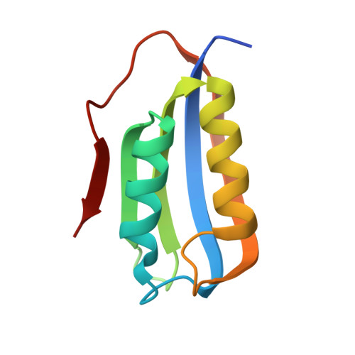
6CBU

Crystal structure of C4S3: A computationally designed immunogen to target Carbohydrate-Occluded Epitopes on the HIV envelope


Experimental Data Snapshot

  • Method: X-RAY DIFFRACTION
  • Resolution: 1.20 Å
  • R-Value Free: 0.153 
  • R-Value Work: 0.143 
  • R-Value Observed: 0.143 

Starting Model: experimental
View more details

wwPDB Validation   3D Report Full Report


This is version 1.4 of the entry. See complete history


Literature

Rationally designed carbohydrate-occluded epitopes elicit HIV-1 Env-specific antibodies.

Zhu, C.Dukhovlinova, E.Council, O.Ping, L.Faison, E.M.Prabhu, S.S.Potter, E.L.Upton, S.L.Yin, G.Fay, J.M.Kincer, L.P.Spielvogel, E.Campbell, S.L.Benhabbour, S.R.Ke, H.Swanstrom, R.Dokholyan, N.V.

(2019) Nat Commun 10: 948-948

  • DOI: https://doi.org/10.1038/s41467-019-08876-w
  • Primary Citation of Related Structures:  
    6CBU, 6CFE

  • PubMed Abstract: 

    An array of carbohydrates masks the HIV-1 surface protein Env, contributing to the evasion of humoral immunity. In most HIV-1 isolates 'glycan holes' occur due to natural sequence variation, potentially revealing the underlying protein surface to the immune system. Here we computationally design epitopes that mimic such surface features (carbohydrate-occluded neutralization epitopes or CONE) of Env through 'epitope transplantation', in which the target region is presented on a carrier protein scaffold with preserved structural properties. Scaffolds displaying the four CONEs are examined for structure and immunogenicity. Crystal structures of two designed proteins reflect the computational models and accurately mimic the native conformations of CONEs. The sera from rabbits immunized with several CONE immunogens display Env binding activity. Our method determines essential structural elements for targets of protective antibodies. The ability to design immunogens with high mimicry to viral proteins also makes possible the exploration of new templates for vaccine development.


  • Organizational Affiliation

    Department of Biochemistry and Biophysics, University of North Carolina at Chapel Hill, Chapel Hill, NC, 27599, USA.


Macromolecules
Find similar proteins by:  (by identity cutoff)  |  3D Structure
Entity ID: 1
MoleculeChains Sequence LengthOrganismDetailsImage
Acylphosphatase-1100Homo sapiensMutation(s): 3 
Gene Names: ACYP1ACYPE
EC: 3.6.1.7
UniProt & NIH Common Fund Data Resources
Find proteins for P07311 (Homo sapiens)
Explore P07311 
Go to UniProtKB:  P07311
PHAROS:  P07311
GTEx:  ENSG00000119640 
Entity Groups  
Sequence Clusters30% Identity50% Identity70% Identity90% Identity95% Identity100% Identity
UniProt GroupP07311
Sequence Annotations
Expand
  • Reference Sequence
Small Molecules
Ligands 1 Unique
IDChains Name / Formula / InChI Key2D Diagram3D Interactions
SO4
Query on SO4

Download Ideal Coordinates CCD File 
B [auth A]SULFATE ION
O4 S
QAOWNCQODCNURD-UHFFFAOYSA-L
Experimental Data & Validation

Experimental Data

  • Method: X-RAY DIFFRACTION
  • Resolution: 1.20 Å
  • R-Value Free: 0.153 
  • R-Value Work: 0.143 
  • R-Value Observed: 0.143 
  • Space Group: P 21 21 21
Unit Cell:
Length ( Å )Angle ( ˚ )
a = 26.069α = 90
b = 57.606β = 90
c = 62.94γ = 90
Software Package:
Software NamePurpose
REFMACrefinement
HKL-2000data scaling
PDB_EXTRACTdata extraction
HKL-2000data reduction
PHASERphasing

Structure Validation

View Full Validation Report


Entry History & Funding Information

Deposition Data


Funding OrganizationLocationGrant Number
National Institutes of Health/National Institute Of Allergy and Infectious Diseases (NIH/NIAID)United StatesR01AI102732

Revision History  (Full details and data files)

  • Version 1.0: 2019-02-06
    Type: Initial release
  • Version 1.1: 2019-02-20
    Changes: Author supporting evidence, Data collection
  • Version 1.2: 2019-03-13
    Changes: Data collection, Database references
  • Version 1.3: 2019-12-18
    Changes: Author supporting evidence
  • Version 1.4: 2023-10-04
    Changes: Data collection, Database references, Refinement description