6C9W | pdb_00006c9w

Crystal Structure of a ligand bound LacY/Nanobody Complex


Experimental Data Snapshot

  • Method: X-RAY DIFFRACTION
  • Resolution: 3.00 Å
  • R-Value Free: 
    0.295 (Depositor), 0.300 (DCC) 
  • R-Value Work: 
    0.276 (Depositor), 0.280 (DCC) 
  • R-Value Observed: 
    0.277 (Depositor) 

Starting Model: experimental
View more details

wwPDB Validation   3D Report Full Report


Ligand Structure Quality Assessment 

Created with Raphaël 2.3.0Worse 01 BetterLigand structure goodness of fit to experimental dataBest fitted 9PGClick on this verticalbar to view details

This is version 2.2 of the entry. See complete history


Literature

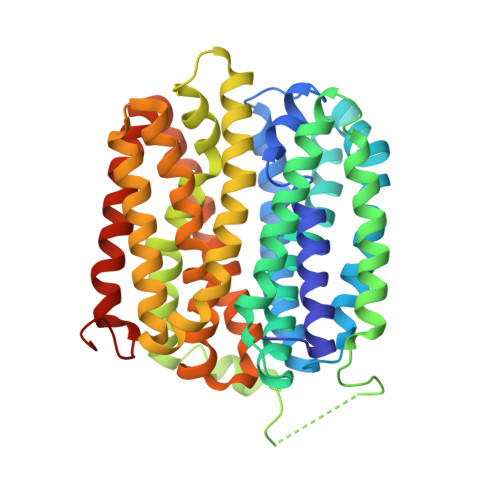
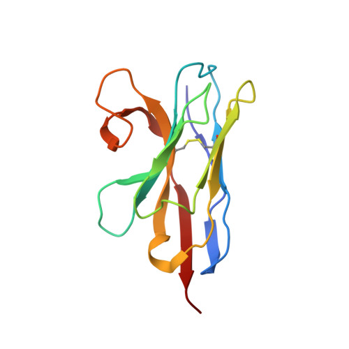
Crystal Structure of a ligand-bound LacY-Nanobody Complex.

Kumar, H.Finer-Moore, J.S.Jiang, X.Smirnova, I.Kasho, V.Pardon, E.Steyaert, J.Kaback, H.R.Stroud, R.M.

(2018) Proc Natl Acad Sci U S A 115: 8769-8774

  • DOI: https://doi.org/10.1073/pnas.1801774115
  • Primary Citation of Related Structures:  
    6C9W

  • PubMed Abstract: 

    The lactose permease of Escherichia coli (LacY), a dynamic polytopic membrane transport protein, catalyzes galactoside/H + symport and operates by an alternating access mechanism that exhibits multiple conformations, the distribution of which is altered by sugar-binding. Camelid nanobodies were made against a double-mutant Gly46 → Trp/Gly262 → Trp (LacY WW ) that produces an outward-open conformation, as opposed to the cytoplasmic open-state crystal structure of WT LacY. Nanobody 9047 (Nb9047) stabilizes WT LacY in a periplasmic-open conformation. Here, we describe the X-ray crystal structure of a complex between LacY WW , the high-affinity substrate analog 4-nitrophenyl-α-d-galactoside (NPG), and Nb9047 at 3-Å resolution. The present crystal structure demonstrates that Nb9047 binds to the periplasmic face of LacY, primarily to the C-terminal six-helical bundle, while a flexible loop of the Nb forms a bridge between the N- and C-terminal halves of LacY across the periplasmic vestibule. The bound Nb partially covers the vestibule, yet does not affect the on-rates or off-rates for the substrate binding to LacY WW , which implicates dynamic flexibility of the Nb-LacY WW complex. Nb9047-binding neither changes the overall structure of LacY WW with bound NPG, nor the positions of side chains comprising the galactoside-binding site. The current NPG-bound structure exhibits a more occluded periplasmic vestibule than seen in a previous structure of a (different Nb) apo-LacY WW /Nb9039 complex that we argue is caused by sugar-binding, with major differences located at the periplasmic ends of transmembrane helices in the N-terminal half of LacY.


  • Organizational Affiliation

    Department of Biochemistry and Biophysics, University of California, San Francisco, CA 94158.


Macromolecules
Find similar proteins by:  (by identity cutoff)  |  3D Structure
Entity ID: 1
MoleculeChains Sequence LengthOrganismDetailsImage
Lactose permease417Escherichia coli K-12Mutation(s): 2 
Gene Names: lacYb0343JW0334
Membrane Entity: Yes 
UniProt
Find proteins for P02920 (Escherichia coli (strain K12))
Explore P02920 
Go to UniProtKB:  P02920
Entity Groups  
Sequence Clusters30% Identity50% Identity70% Identity90% Identity95% Identity100% Identity
UniProt GroupP02920
Sequence Annotations
Expand
  • Reference Sequence
Find similar proteins by:  (by identity cutoff)  |  3D Structure
Entity ID: 2
MoleculeChains Sequence LengthOrganismDetailsImage
Nanobody9047125Lama glamaMutation(s): 0 
Entity Groups  
Sequence Clusters30% Identity50% Identity70% Identity90% Identity95% Identity100% Identity
Sequence Annotations
Expand
  • Reference Sequence
Experimental Data & Validation

Experimental Data

  • Method: X-RAY DIFFRACTION
  • Resolution: 3.00 Å
  • R-Value Free:  0.295 (Depositor), 0.300 (DCC) 
  • R-Value Work:  0.276 (Depositor), 0.280 (DCC) 
  • R-Value Observed: 0.277 (Depositor) 
Space Group: P 65 2 2
Unit Cell:
Length ( Å )Angle ( ˚ )
a = 153.55α = 90
b = 153.55β = 90
c = 193.68γ = 120
Software Package:
Software NamePurpose
PHENIXrefinement
XSCALEdata scaling
PDB_EXTRACTdata extraction
XDSdata reduction
PHENIXphasing

Structure Validation

View Full Validation Report


Ligand Structure Quality Assessment 

Created with Raphaël 2.3.0Worse 01 BetterLigand structure goodness of fit to experimental dataBest fitted 9PGClick on this verticalbar to view details

Entry History & Funding Information

Deposition Data


Funding OrganizationLocationGrant Number
National Institutes of Health/National Institute of General Medical Sciences (NIH/NIGMS)United StatesGM120043

Revision History  (Full details and data files)

  • Version 1.0: 2018-08-15
    Type: Initial release
  • Version 1.1: 2018-08-29
    Changes: Data collection, Database references
  • Version 1.2: 2018-09-05
    Changes: Data collection, Database references
  • Version 1.3: 2019-02-20
    Changes: Author supporting evidence, Data collection
  • Version 1.4: 2020-01-01
    Changes: Author supporting evidence
  • Version 2.0: 2020-07-29
    Type: Remediation
    Reason: Carbohydrate remediation
    Changes: Atomic model, Data collection, Derived calculations, Structure summary
  • Version 2.1: 2023-10-04
    Changes: Data collection, Database references, Refinement description, Structure summary
  • Version 2.2: 2024-10-16
    Changes: Structure summary