


The A225L Substitution of hUGDH alters structure and function
Gross, P.G., Wood, Z.A.To be published.
Experimental Data Snapshot
Starting Model: experimental
View more details
Entity ID: 1 | |||||
|---|---|---|---|---|---|
| Molecule | Chains | Sequence Length | Organism | Details | Image |
| UDP-glucose 6-dehydrogenase | 494 | Homo sapiens | Mutation(s): 1 Gene Names: UGDH EC: 1.1.1.22 | 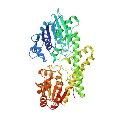 | |
UniProt & NIH Common Fund Data Resources | |||||
PHAROS: O60701 GTEx: ENSG00000109814 | |||||
Entity Groups | |||||
| Sequence Clusters | 30% Identity50% Identity70% Identity90% Identity95% Identity100% Identity | ||||
| UniProt Group | O60701 | ||||
Sequence AnnotationsExpand | |||||
Reference Sequence | |||||
| Ligands 2 Unique | |||||
|---|---|---|---|---|---|
| ID | Chains | Name / Formula / InChI Key | 2D Diagram | 3D Interactions | |
| UDX Download:Ideal Coordinates CCD File | G [auth A] H [auth A] J [auth B] K [auth B] M [auth C] | URIDINE-5'-DIPHOSPHATE-XYLOPYRANOSE C14 H22 N2 O16 P2 DQQDLYVHOTZLOR-OCIMBMBZSA-N |  | ||
| CL Download:Ideal Coordinates CCD File | I [auth A], L [auth B], P [auth D] | CHLORIDE ION Cl VEXZGXHMUGYJMC-UHFFFAOYSA-M |  | ||
| Length ( Å ) | Angle ( ˚ ) |
|---|---|
| a = 93.52 | α = 90 |
| b = 194.99 | β = 110.71 |
| c = 109.54 | γ = 90 |
| Software Name | Purpose |
|---|---|
| PHENIX | refinement |
| XDS | data reduction |
| XSCALE | data scaling |
| PHASER | phasing |
| Coot | model building |
| Funding Organization | Location | Grant Number |
|---|---|---|
| National Institutes of Health/National Institute of General Medical Sciences (NIH/NIGMS) | United States | R01GM114298 |


