6C2Z | pdb_00006c2z

Crystal Structures of Cystathionine beta-Synthase from Saccharomyces cerevisiae: the Structure of the PLP-Aminoacrylate Intermediate


Experimental Data Snapshot

  • Method: X-RAY DIFFRACTION
  • Resolution: 1.37 Å
  • R-Value Free: 
    0.163 (Depositor), 0.163 (DCC) 
  • R-Value Work: 
    0.134 (Depositor), 0.134 (DCC) 
  • R-Value Observed: 
    0.136 (Depositor) 

Starting Model: experimental
View more details

wwPDB Validation 3D Report Full Report

Validation slider image for 6C2Z

Ligand Structure Quality Assessment 


This is version 1.4 of the entry. See complete history

Literature

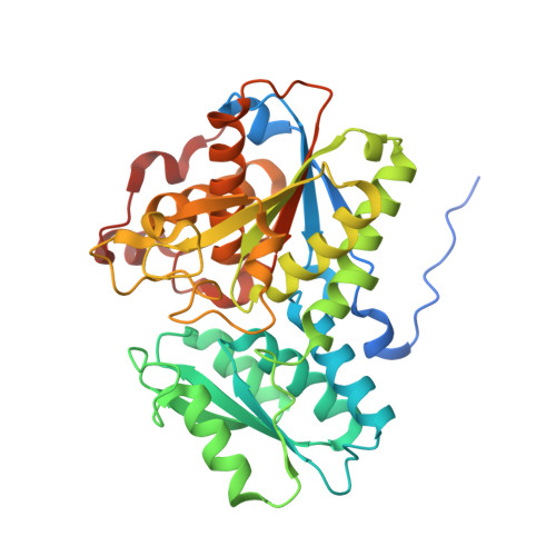
Crystal Structures of Cystathionine beta-Synthase from Saccharomyces cerevisiae: One Enzymatic Step at a Time.

Tu, Y.Kreinbring, C.A.Hill, M.Liu, C.Petsko, G.A.McCune, C.D.Berkowitz, D.B.Liu, D.Ringe, D.

(2018) Biochemistry 57: 3134-3145

  • DOI: https://doi.org/10.1021/acs.biochem.8b00092
  • Primary Citation Related Structures: 
    6C2H, 6C2Q, 6C2Z, 6C4P

  • PubMed Abstract: 

    Cystathionine β-synthase (CBS) is a key regulator of sulfur amino acid metabolism, taking homocysteine from the methionine cycle to the biosynthesis of cysteine via the trans-sulfuration pathway. CBS is also a predominant source of H 2 S biogenesis. Roles for CBS have been reported for neuronal death pursuant to cerebral ischemia, promoting ovarian tumor growth, and maintaining drug-resistant phenotype by controlling redox behavior and regulating mitochondrial bioenergetics. The trans-sulfuration pathway is well-conserved in eukaryotes, but the analogous enzymes have different enzymatic behavior in different organisms. CBSs from the higher organisms contain a heme in an N-terminal domain. Though the presence of the heme, whose functions in CBSs have yet to be elucidated, is biochemically interesting, it hampers UV-vis absorption spectroscopy investigations of pyridoxal 5'-phosphate (PLP) species. CBS from Saccharomyces cerevisiae (yCBS) naturally lacks the heme-containing N-terminal domain, which makes it an ideal model for spectroscopic studies of the enzymological reaction catalyzed and allows structural studies of the basic yCBS catalytic core (yCBS-cc). Here we present the crystal structure of yCBS-cc, solved to 1.5 Å. Crystal structures of yCBS-cc in complex with enzymatic reaction intermediates have been captured, providing a structural basis for residues involved in catalysis. Finally, the structure of the yCBS-cc cofactor complex generated by incubation with an inhibitor shows apparent off-pathway chemistry not normally seen with CBS.


  • Organizational Affiliation
    • Department of Biochemistry , Brandeis University , Waltham , Massachusetts 02454 , United States.

Macromolecule Content 

  • Total Structure Weight: 42.5 kDa 
  • Atom Count: 3,263 
  • Modeled Residue Count: 345 
  • Deposited Residue Count: 375 
  • Unique protein chains: 1

Macromolecules

Find similar proteins by:|  3D Structure
Entity ID: 1
MoleculeChains  Sequence LengthOrganismDetailsImage
Cystathionine beta-synthase375Saccharomyces cerevisiae S288CMutation(s): 0 
Gene Names: CYS4STR4YGR155WG6667
EC: 4.2.1.22
UniProt
Find proteins for P32582 (Saccharomyces cerevisiae (strain ATCC 204508 / S288c))
Explore P32582 
Go to UniProtKB:  P32582
Entity Groups
Sequence Clusters30% Identity50% Identity70% Identity90% Identity95% Identity100% Identity
UniProt GroupP32582
Sequence Annotations
Expand
Reference Sequence

Small Molecules

Ligands 7 Unique
IDChains Name / Formula / InChI Key2D Diagram3D Interactions
P1T

Query on P1T


Download:Ideal Coordinates CCD File
B [auth A]2-[({3-HYDROXY-2-METHYL-5-[(PHOSPHONOOXY)METHYL]PYRIDIN-4-YL}METHYL)AMINO]ACRYLIC ACID
C11 H15 N2 O7 P
BXUDKFHCAMQSRX-UHFFFAOYSA-N
PGE

Query on PGE


Download:Ideal Coordinates CCD File
H [auth A]TRIETHYLENE GLYCOL
C6 H14 O4
ZIBGPFATKBEMQZ-UHFFFAOYSA-N
PEG

Query on PEG


Download:Ideal Coordinates CCD File
F [auth A],
G [auth A]
DI(HYDROXYETHYL)ETHER
C4 H10 O3
MTHSVFCYNBDYFN-UHFFFAOYSA-N
EDO

Query on EDO


Download:Ideal Coordinates CCD File
I [auth A]
J [auth A]
K [auth A]
L [auth A]
M [auth A]
I [auth A],
J [auth A],
K [auth A],
L [auth A],
M [auth A],
N [auth A],
O [auth A]
1,2-ETHANEDIOL
C2 H6 O2
LYCAIKOWRPUZTN-UHFFFAOYSA-N
CA

Query on CA


Download:Ideal Coordinates CCD File
C [auth A]CALCIUM ION
Ca
BHPQYMZQTOCNFJ-UHFFFAOYSA-N
CL

Query on CL


Download:Ideal Coordinates CCD File
E [auth A]CHLORIDE ION
Cl
VEXZGXHMUGYJMC-UHFFFAOYSA-M
NA

Query on NA


Download:Ideal Coordinates CCD File
D [auth A]SODIUM ION
Na
FKNQFGJONOIPTF-UHFFFAOYSA-N

Experimental Data & Validation

Experimental Data

  • Method: X-RAY DIFFRACTION
  • Resolution: 1.37 Å
  • R-Value Free:  0.163 (Depositor), 0.163 (DCC) 
  • R-Value Work:  0.134 (Depositor), 0.134 (DCC) 
  • R-Value Observed: 0.136 (Depositor) 
Space Group: P 65 2 2
Unit Cell:
Length ( Å )Angle ( ˚ )
a = 81.465α = 90
b = 81.465β = 90
c = 209.486γ = 120
Software Package:
Software NamePurpose
REFMACrefinement
PHENIXphasing
HKL-2000data reduction
SCALEPACKdata scaling
PDB_EXTRACTdata extraction

Structure Validation

View Full Validation Report


Ligand Structure Quality Assessment 


Entry History 

& Funding Information

Deposition Data


Funding OrganizationLocationGrant Number
National Institutes of Health/National Institute of General Medical Sciences (NIH/NIGMS)United StatesGM32415

Revision History  (Full details and data files)

  • Version 1.0: 2018-04-25
    Type: Initial release
  • Version 1.1: 2018-06-20
    Changes: Data collection, Database references, Derived calculations
  • Version 1.2: 2019-02-20
    Changes: Author supporting evidence, Data collection
  • Version 1.3: 2020-01-01
    Changes: Author supporting evidence
  • Version 1.4: 2023-10-04
    Changes: Data collection, Database references, Derived calculations, Refinement description