6BTK | pdb_00006btk

Segment from bank vole prion protein 168-176 QYNNQNNFV


Experimental Data Snapshot

  • Method: X-RAY DIFFRACTION
  • Resolution: 1.10 Å
  • R-Value Free: 
    0.162 (Depositor), 0.171 (DCC) 
  • R-Value Work: 
    0.139 (Depositor), 0.140 (DCC) 
  • R-Value Observed: 
    0.141 (Depositor) 

wwPDB Validation 3D Report Full Report

Validation slider image for 6BTK

This is version 1.3 of the entry. See complete history

Literature

Sub-angstrom cryo-EM structure of a prion protofibril reveals a polar clasp.

Gallagher-Jones, M.Glynn, C.Boyer, D.R.Martynowycz, M.W.Hernandez, E.Miao, J.Zee, C.T.Novikova, I.V.Goldschmidt, L.McFarlane, H.T.Helguera, G.F.Evans, J.E.Sawaya, M.R.Cascio, D.Eisenberg, D.S.Gonen, T.Rodriguez, J.A.

(2018) Nat Struct Mol Biol 25: 131-134

  • DOI: https://doi.org/10.1038/s41594-017-0018-0
  • Primary Citation Related Structures: 
    6AXZ, 6BTK

  • PubMed Abstract: 

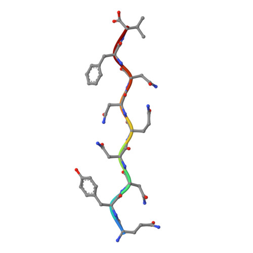
    The atomic structure of the infectious, protease-resistant, β-sheet-rich and fibrillar mammalian prion remains unknown. Through the cryo-EM method MicroED, we reveal the sub-ångström-resolution structure of a protofibril formed by a wild-type segment from the β2-α2 loop of the bank vole prion protein. The structure of this protofibril reveals a stabilizing network of hydrogen bonds that link polar zippers within a sheet, producing motifs we have named 'polar clasps'.


  • Organizational Affiliation
    • Department of Chemistry and Biochemistry, UCLA-DOE Institute for Genomics and Proteomics, University of California Los Angeles, Los Angeles, CA, USA.

Macromolecule Content 

  • Total Structure Weight: 1.14 kDa 
  • Atom Count: 85 
  • Modeled Residue Count: 9 
  • Deposited Residue Count: 9 
  • Unique protein chains: 1

Macromolecules

Find similar proteins by:  Sequence   |   3D Structure  
Entity ID: 1
MoleculeChains  Sequence LengthOrganismDetailsImage
Major prion protein9Myodes glareolusMutation(s): 0 
UniProt
Find proteins for Q8VHV5 (Myodes glareolus)
Explore Q8VHV5 
Go to UniProtKB:  Q8VHV5
Entity Groups
UniProt GroupQ8VHV5
Sequence Annotations
Expand
Reference Sequence

Experimental Data & Validation

Experimental Data

  • Method: X-RAY DIFFRACTION
  • Resolution: 1.10 Å
  • R-Value Free:  0.162 (Depositor), 0.171 (DCC) 
  • R-Value Work:  0.139 (Depositor), 0.140 (DCC) 
  • R-Value Observed: 0.141 (Depositor) 
Space Group: P 1
Unit Cell:
Length ( Å )Angle ( ˚ )
a = 4.874α = 93.36
b = 10.107β = 91.15
c = 30.415γ = 101.66
Software Package:
Software NamePurpose
REFMACrefinement
HKL-2000data scaling
PHASERphasing
PDB_EXTRACTdata extraction
HKL-2000data reduction

Structure Validation

View Full Validation Report


Entry History 

Deposition Data

Revision History  (Full details and data files)

  • Version 1.0: 2018-01-17
    Type: Initial release
  • Version 1.1: 2018-01-31
    Changes: Database references
  • Version 1.2: 2018-02-21
    Changes: Database references
  • Version 1.3: 2024-03-13
    Changes: Data collection, Database references