6BTG | pdb_00006btg

Crystal structure of deoxyribose-phosphate aldolase bound with DHAP from Bacillus Thuringiensis


Experimental Data Snapshot

  • Method: X-RAY DIFFRACTION
  • Resolution: 1.70 Å
  • R-Value Free: 
    0.225 (Depositor), 0.225 (DCC) 
  • R-Value Work: 
    0.186 (Depositor), 0.187 (DCC) 
  • R-Value Observed: 
    0.188 (Depositor) 

Starting Model: experimental
View more details

wwPDB Validation 3D Report Full Report

Validation slider image for 6BTG

Ligand Structure Quality Assessment 


This is version 1.2 of the entry. See complete history

Literature

Salvage of the 5-deoxyribose byproduct of radical SAM enzymes.

Beaudoin, G.A.W.Li, Q.Folz, J.Fiehn, O.Goodsell, J.L.Angerhofer, A.Bruner, S.D.Hanson, A.D.

(2018) Nat Commun 9: 3105-3105

  • DOI: https://doi.org/10.1038/s41467-018-05589-4
  • Primary Citation Related Structures: 
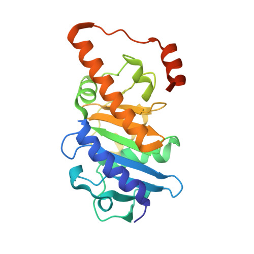
    6BTD, 6BTG

  • PubMed Abstract: 

    5-Deoxyribose is formed from 5'-deoxyadenosine, a toxic byproduct of radical S-adenosylmethionine (SAM) enzymes. The degradative fate of 5-deoxyribose is unknown. Here, we define a salvage pathway for 5-deoxyribose in bacteria, consisting of phosphorylation, isomerization, and aldol cleavage steps. Analysis of bacterial genomes uncovers widespread, unassigned three-gene clusters specifying a putative kinase, isomerase, and sugar phosphate aldolase. We show that the enzymes encoded by the Bacillus thuringiensis cluster, acting together in vitro, convert 5-deoxyribose successively to 5-deoxyribose 1-phosphate, 5-deoxyribulose 1-phosphate, and dihydroxyacetone phosphate plus acetaldehyde. Deleting the isomerase decreases the 5-deoxyribulose 1-phosphate pool size, and deleting either the isomerase or the aldolase increases susceptibility to 5-deoxyribose. The substrate preference of the aldolase is unique among family members, and the X-ray structure reveals an unusual manganese-dependent enzyme. This work defines a salvage pathway for 5-deoxyribose, a near-universal metabolite.


  • Organizational Affiliation
    • Horticultural Sciences Department, University of Florida, Gainesville, FL, 32611, USA.

Macromolecule Content 

  • Total Structure Weight: 25.17 kDa 
  • Atom Count: 1,886 
  • Modeled Residue Count: 207 
  • Deposited Residue Count: 222 
  • Unique protein chains: 1

Macromolecules

Find similar proteins by:|  3D Structure
Entity ID: 1
MoleculeChains  Sequence LengthOrganismDetailsImage
Fuculose phosphate aldolase222Bacillus thuringiensisMutation(s): 0 
Gene Names: BK775_23715CCZ40_07290
EC: 4.1.2.17

Small Molecules

Experimental Data & Validation

Experimental Data

  • Method: X-RAY DIFFRACTION
  • Resolution: 1.70 Å
  • R-Value Free:  0.225 (Depositor), 0.225 (DCC) 
  • R-Value Work:  0.186 (Depositor), 0.187 (DCC) 
  • R-Value Observed: 0.188 (Depositor) 
Space Group: P 4 21 2
Unit Cell:
Length ( Å )Angle ( ˚ )
a = 105.104α = 90
b = 105.104β = 90
c = 49.366γ = 90
Software Package:
Software NamePurpose
PHENIXrefinement
XDSdata reduction
XDSdata scaling
PHASERphasing

Structure Validation

View Full Validation Report


Ligand Structure Quality Assessment 


Entry History 

Deposition Data

Revision History  (Full details and data files)

  • Version 1.0: 2018-07-04
    Type: Initial release
  • Version 1.1: 2019-05-08
    Changes: Data collection, Database references
  • Version 1.2: 2023-10-04
    Changes: Data collection, Database references, Refinement description