6BR6 | pdb_00006br6

N2 neuraminidase in complex with a novel antiviral compound


Experimental Data Snapshot

  • Method: X-RAY DIFFRACTION
  • Resolution: 2.04 Å
  • R-Value Free: 
    0.225 (Depositor), 0.227 (DCC) 
  • R-Value Work: 
    0.173 (Depositor), 0.174 (DCC) 
  • R-Value Observed: 
    0.176 (Depositor) 

wwPDB Validation 3D Report Full Report

Validation slider image for 6BR6

Ligand Structure Quality Assessment 


This is version 3.1 of the entry. See complete history. 

Literature

A Sulfonozanamivir Analogue Has Potent Anti-influenza Virus Activity.

Hadhazi, A., Li, L., Bailly, B., Maggioni, A., Martin, G., Dirr, L., Dyason, J.C., Thomson, R.J., Gao, G.F., Borbas, A., Ve, T., Pascolutti, M., von Itzstein, M.

(2018) ChemMedChem 13: 785-789

  • DOI: https://doi.org/10.1002/cmdc.201800092
  • Primary Citation Related Structures: 
    6BR5, 6BR6

  • PubMed Abstract: 

    Influenza virus infection continues to cause significant, often severe, respiratory illness worldwide. A validated target for the development of anti-influenza agents is the virus surface protein sialidase. In the current study, we have discovered a highly potent inhibitor of influenza virus sialidase, based on a novel sialosyl sulfonate template. The synthesised 3-guanidino sialosyl α-sulfonate, a sulfonozanamivir analogue, inhibits viral replication in vitro at the nanomolar level, comparable to that of the anti-influenza drug zanamivir. Using protein X-ray crystallography we show that the sialosyl α-sulfonate template binds within the sialidase active site in a 1 C 4 chair conformation. The C1-sulfonate moiety forms crucial and strong-binding interactions with the active site's triarginyl cluster, while the 3-guanidino moiety interacts significantly with conserved active site residues. This sulfonozanamivir analogue provides a new direction in anti-influenza virus drug development.


  • Organizational Affiliation: 
    • Institute for Glycomics, Gold Coast Campus, Griffith University, Queensland, 4222, Australia.

Macromolecule Content 

  • Total Structure Weight: 44.61 kDa 
  • Atom Count: 3,289 
  • Modeled Residue Count: 387 
  • Deposited Residue Count: 387 
  • Unique protein chains: 1

Macromolecules

Find similar proteins by:|  3D Structure
Entity ID: 1
MoleculeChains  Sequence LengthOrganismDetailsImage
Neuraminidase387Influenza A virus (A/Perth/16/2009(H3N2))Mutation(s): 0 
Gene Names: NA
EC: 3.2.1.18
UniProt
Find proteins for E0UY46 (Influenza A virus)
Explore E0UY46 
Go to UniProtKB:  E0UY46
Entity Groups
Sequence Clusters30% Identity50% Identity70% Identity90% Identity95% Identity100% Identity
UniProt GroupE0UY46
Glycosylation
Glycosylation Sites: 2
Sequence Annotations
Expand
Reference Sequence

Oligosaccharides

Help  
Entity ID: 2
MoleculeChains Length2D Diagram GlycosylationD Interactions
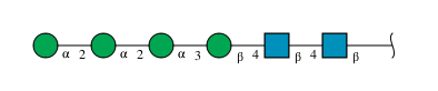
alpha-D-mannopyranose-(1-2)-alpha-D-mannopyranose-(1-2)-alpha-D-mannopyranose-(1-3)-beta-D-mannopyranose-(1-4)-2-acetamido-2-deoxy-beta-D-glucopyranose-(1-4)-2-acetamido-2-deoxy-beta-D-glucopyranose
B
6N-Glycosylation
Glycosylation Resources
GlyTouCan: G01760ZU
GlyCosmos: G01760ZU
GlyGen: G01760ZU

Small Molecules

Ligands 5 Unique
IDChains Name / Formula / InChI Key2D Diagram3D Interactions
E3M

Query on E3M


Download:Ideal Coordinates CCD File
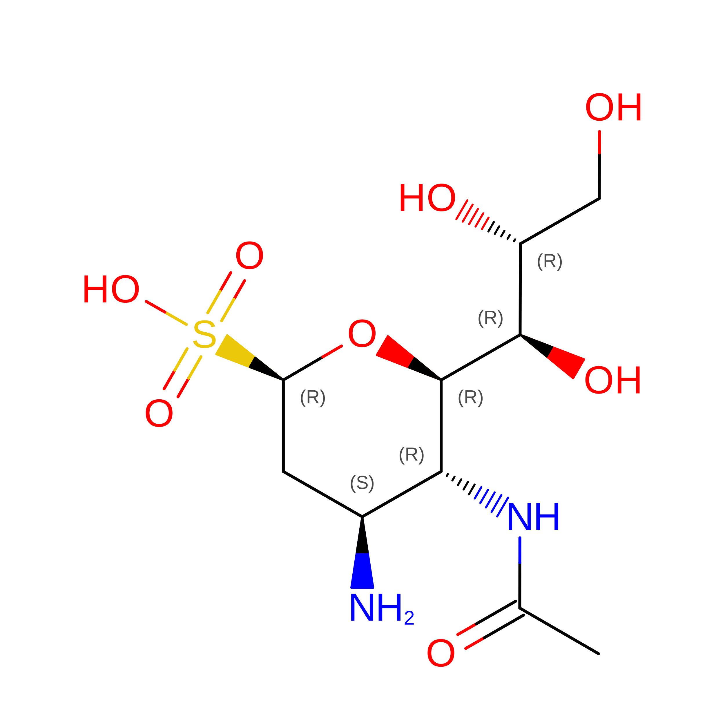
G [auth A](1R)-4-acetamido-3-amino-1,5-anhydro-2,3,4-trideoxy-1-sulfo-D-glycero-D-galacto-octitol
C10 H20 N2 O8 S
XAZGDFCTWHWJMA-IHICSVBISA-N
NAG

Query on NAG


Download:Ideal Coordinates CCD File
C [auth A]2-acetamido-2-deoxy-beta-D-glucopyranose
C8 H15 N O6
OVRNDRQMDRJTHS-FMDGEEDCSA-N
GOL

Query on GOL


Download:Ideal Coordinates CCD File
F [auth A]GLYCEROL
C3 H8 O3
PEDCQBHIVMGVHV-UHFFFAOYSA-N
NI

Query on NI


Download:Ideal Coordinates CCD File
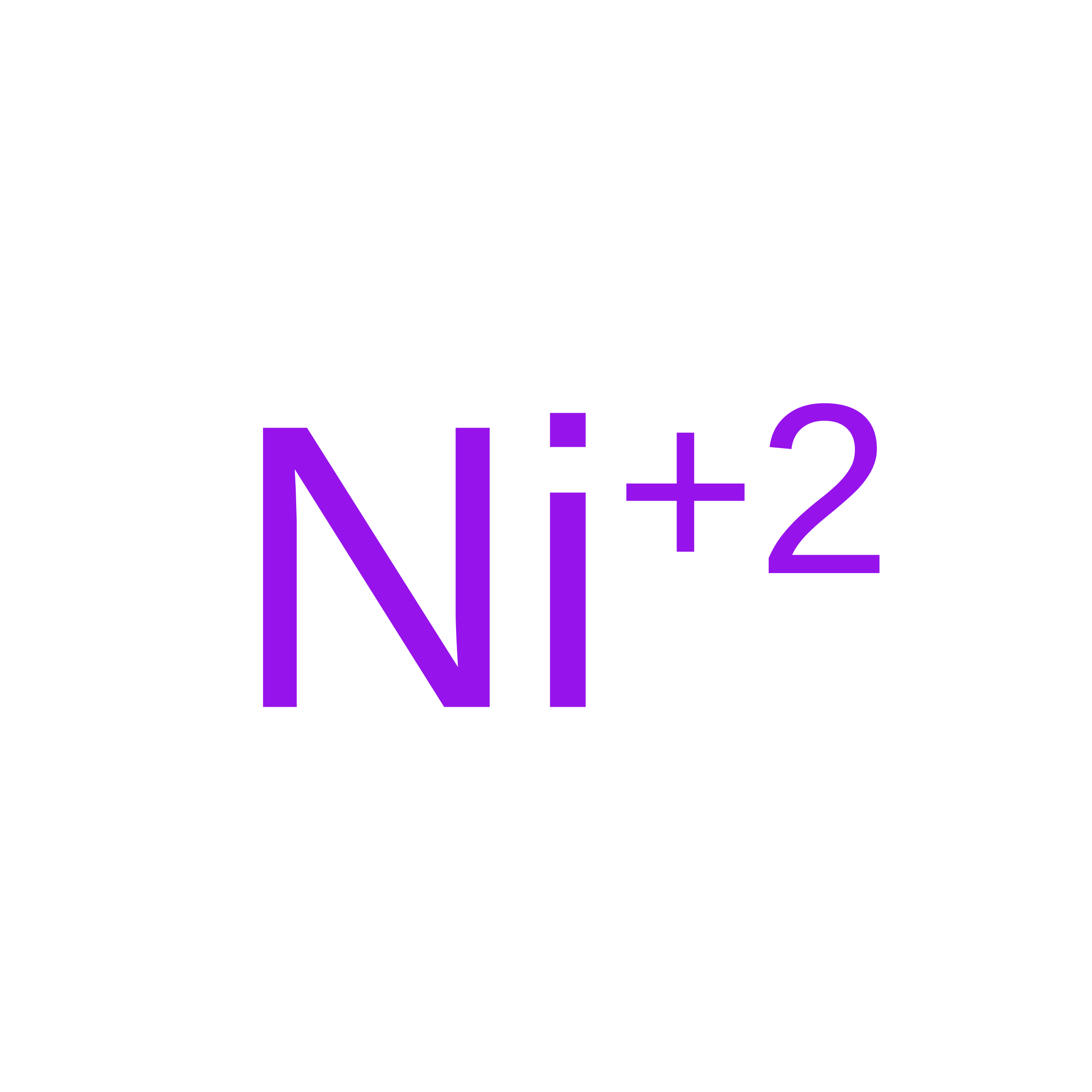
D [auth A]NICKEL (II) ION
Ni
VEQPNABPJHWNSG-UHFFFAOYSA-N
CA

Query on CA


Download:Ideal Coordinates CCD File
E [auth A]CALCIUM ION
Ca
BHPQYMZQTOCNFJ-UHFFFAOYSA-N

Experimental Data & Validation

Experimental Data

  • Method: X-RAY DIFFRACTION
  • Resolution: 2.04 Å
  • R-Value Free:  0.225 (Depositor), 0.227 (DCC) 
  • R-Value Work:  0.173 (Depositor), 0.174 (DCC) 
  • R-Value Observed: 0.176 (Depositor) 
Space Group: P 4 21 2
Unit Cell:
Length ( Å )Angle ( ˚ )
a = 108.457α = 90
b = 108.457β = 90
c = 70.204γ = 90
Software Package:
Software NamePurpose
PHENIXrefinement
XDSdata reduction
Aimlessdata scaling
PHENIXphasing

Structure Validation

View Full Validation Report


Ligand Structure Quality Assessment 


Entry History 

& Funding Information

Deposition Data


Funding OrganizationLocationGrant Number
National Health and Medical Research Council (NHMRC, Australia)Australia1071659

Revision History  (Full details and data files)

  • Version 1.0: 2018-03-07
    Type: Initial release
  • Version 1.1: 2018-03-14
    Changes: Database references, Source and taxonomy
  • Version 1.2: 2018-05-09
    Changes: Data collection, Database references
  • Version 1.3: 2019-02-20
    Changes: Author supporting evidence, Data collection
  • Version 1.4: 2020-01-08
    Changes: Author supporting evidence
  • Version 2.0: 2020-05-13
    Changes: Atomic model, Data collection
  • Version 3.0: 2020-07-29
    Type: Remediation
    Reason: Carbohydrate remediation
    Changes: Atomic model, Data collection, Derived calculations, Structure summary
  • Version 3.1: 2024-10-09
    Changes: Data collection, Database references, Derived calculations, Structure summary