6BQC | pdb_00006bqc

Cyclopropane fatty acid synthase from E. coli

  • Classification: TRANSFERASE
  • Organism(s): Escherichia coli
  • Expression System: Escherichia coli
  • Mutation(s): No 

  • Deposited: 2017-11-27 Released: 2018-07-04 
  • Deposition Author(s): Hari, S.B., Grant, R.A., Sauer, R.T.
  • Funding Organization(s): National Institutes of Health/National Institute Of Allergy and Infectious Diseases (NIH/NIAID), National Institutes of Health/National Institute of General Medical Sciences (NIH/NIGMS)

Experimental Data Snapshot

  • Method: X-RAY DIFFRACTION
  • Resolution: 2.07 Å
  • R-Value Free: 
    0.205 (Depositor), 0.208 (DCC) 
  • R-Value Work: 
    0.170 (Depositor), 0.172 (DCC) 
  • R-Value Observed: 
    0.172 (Depositor) 

Starting Model: experimental
View more details

wwPDB Validation 3D Report Full Report

Validation slider image for 6BQC

Ligand Structure Quality Assessment 


This is version 1.5 of the entry. See complete history

Literature

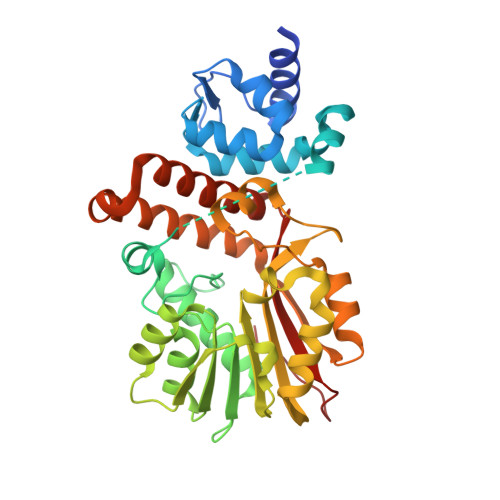
Structural and Functional Analysis of E. coli Cyclopropane Fatty Acid Synthase.

Hari, S.B.Grant, R.A.Sauer, R.T.

(2018) Structure 26: 1251

  • DOI: https://doi.org/10.1016/j.str.2018.06.008
  • Primary Citation Related Structures: 
    6BQC

  • PubMed Abstract: 

    Cell membranes must adapt to different environments. In Gram-negative bacteria, the inner membrane can be remodeled directly by modification of lipids embedded in the bilayer. For example, when Escherichia coli enters stationary phase, cyclopropane fatty acid (CFA) synthase converts most double bonds in unsaturated inner-membrane lipids into cyclopropyl groups. Here we report the crystal structure of E. coli CFA synthase. The enzyme is a dimer in the crystal and in solution, with each subunit containing a smaller N-domain that associates tightly with a larger catalytic C-domain, even following cleavage of the inter-domain linker or co-expression of each individual domain. Efficient catalysis requires dimerization and proper linkage of the two domains. These findings support an avidity-based model in which one subunit of the dimer stabilizes membrane binding, while the other subunit carries out catalysis.


  • Organizational Affiliation
    • Department of Biology, Massachusetts Institute of Technology, Cambridge, MA 02139, USA.

Macromolecule Content 

  • Total Structure Weight: 45.37 kDa 
  • Atom Count: 3,098 
  • Modeled Residue Count: 348 
  • Deposited Residue Count: 383 
  • Unique protein chains: 1

Macromolecules

Find similar proteins by:|  3D Structure
Entity ID: 1
MoleculeChains  Sequence LengthOrganismDetailsImage
Cyclopropane-fatty-acyl-phospholipid synthase383Escherichia coliMutation(s): 0 
Gene Names: cfa
EC: 2.1.1.79
UniProt
Find proteins for P0A9H7 (Escherichia coli (strain K12))
Explore P0A9H7 
Go to UniProtKB:  P0A9H7
Entity Groups
Sequence Clusters30% Identity50% Identity70% Identity90% Identity95% Identity100% Identity
UniProt GroupP0A9H7
Sequence Annotations
Expand
Reference Sequence

Small Molecules

Ligands 3 Unique
IDChains Name / Formula / InChI Key2D Diagram3D Interactions
LOP

Query on LOP


Download:Ideal Coordinates CCD File
C [auth A](1R)-2-{[(R)-(2-AMINOETHOXY)(HYDROXY)PHOSPHORYL]OXY}-1-[(DODECANOYLOXY)METHYL]ETHYL (9Z)-OCTADEC-9-ENOATE
C35 H68 N O8 P
FUUNMZKPCMPCHT-ILGKRYBBSA-N
GOL

Query on GOL


Download:Ideal Coordinates CCD File
D [auth A]
E [auth A]
F [auth A]
G [auth A]
H [auth A]
D [auth A],
E [auth A],
F [auth A],
G [auth A],
H [auth A],
I [auth A],
J [auth A]
GLYCEROL
C3 H8 O3
PEDCQBHIVMGVHV-UHFFFAOYSA-N
CO3

Query on CO3


Download:Ideal Coordinates CCD File
B [auth A]CARBONATE ION
C O3
BVKZGUZCCUSVTD-UHFFFAOYSA-L

Experimental Data & Validation

Experimental Data

  • Method: X-RAY DIFFRACTION
  • Resolution: 2.07 Å
  • R-Value Free:  0.205 (Depositor), 0.208 (DCC) 
  • R-Value Work:  0.170 (Depositor), 0.172 (DCC) 
  • R-Value Observed: 0.172 (Depositor) 
Space Group: C 2 2 21
Unit Cell:
Length ( Å )Angle ( ˚ )
a = 75.425α = 90
b = 102.981β = 90
c = 157.031γ = 90
Software Package:
Software NamePurpose
HKL-2000data collection
HKL-2000data scaling
PHASERphasing
PHENIXrefinement
PDB_EXTRACTdata extraction

Structure Validation

View Full Validation Report


Ligand Structure Quality Assessment 


Entry History 

& Funding Information

Deposition Data


Funding OrganizationLocationGrant Number
National Institutes of Health/National Institute Of Allergy and Infectious Diseases (NIH/NIAID)United StatesR01 AI-016892
National Institutes of Health/National Institute of General Medical Sciences (NIH/NIGMS)United StatesF32GM116241

Revision History  (Full details and data files)

  • Version 1.0: 2018-07-04
    Type: Initial release
  • Version 1.1: 2018-08-15
    Changes: Data collection, Database references
  • Version 1.2: 2018-09-19
    Changes: Data collection, Database references
  • Version 1.3: 2019-02-20
    Changes: Author supporting evidence, Data collection
  • Version 1.4: 2019-12-11
    Changes: Author supporting evidence
  • Version 1.5: 2023-10-04
    Changes: Data collection, Database references, Refinement description