6BP2 | pdb_00006bp2

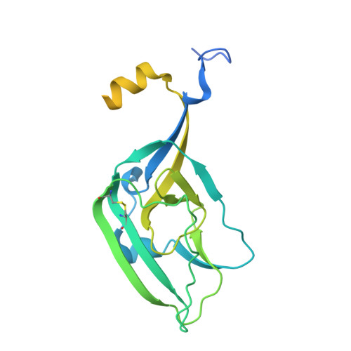
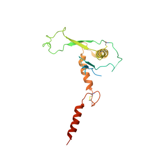
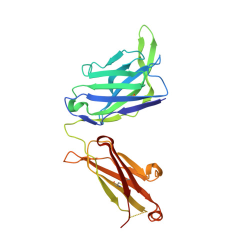
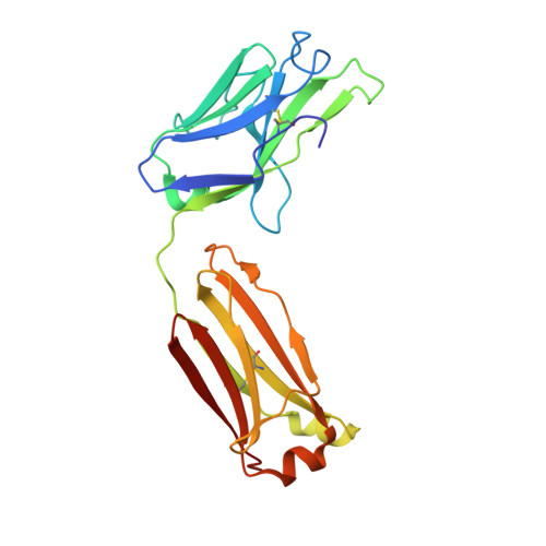
Therapeutic human monoclonal antibody MR191 bound to a marburgvirus glycoprotein


Experimental Data Snapshot

  • Method: X-RAY DIFFRACTION
  • Resolution: 3.17 Å
  • R-Value Free: 
    0.284 (Depositor), 0.285 (DCC) 
  • R-Value Work: 
    0.252 (Depositor), 0.256 (DCC) 
  • R-Value Observed: 
    0.254 (Depositor) 

Starting Model: experimental
View more details

wwPDB Validation 3D Report Full Report

Validation slider image for 6BP2

This is version 3.2 of the entry. See complete history. 

Literature

The Marburgvirus-Neutralizing Human Monoclonal Antibody MR191 Targets a Conserved Site to Block Virus Receptor Binding.

King, L.B., Fusco, M.L., Flyak, A.I., Ilinykh, P.A., Huang, K., Gunn, B., Kirchdoerfer, R.N., Hastie, K.M., Sangha, A.K., Meiler, J., Alter, G., Bukreyev, A., Crowe, J.E., Saphire, E.O.

(2018) Cell Host Microbe 23: 101-109.e4

  • DOI: https://doi.org/10.1016/j.chom.2017.12.003
  • Primary Citation Related Structures: 
    6BP2

  • PubMed Abstract: 

    Since their first identification 50 years ago, marburgviruses have emerged several times, with 83%-90% lethality in the largest outbreaks. Although no vaccines or therapeutics are available for human use, the human antibody MR191 provides complete protection in non-human primates when delivered several days after inoculation of a lethal marburgvirus dose. The detailed neutralization mechanism of MR191 remains outstanding. Here we present a 3.2 Å crystal structure of MR191 complexed with a trimeric marburgvirus surface glycoprotein (GP). MR191 neutralizes by occupying the conserved receptor-binding site and competing with the host receptor Niemann-Pick C1. The structure illuminates previously disordered regions of GP including the stalk, fusion loop, CX 6 CC switch, and an N-terminal region of GP2 that wraps about the outside of GP1 to anchor a marburgvirus-specific "wing" antibody epitope. Virus escape mutations mapped far outside the MR191 receptor-binding site footprint suggest a role for these other regions in the GP quaternary structure.


  • Organizational Affiliation: 
    • Department of Immunology and Microbiology, The Scripps Research Institute, La Jolla, CA 92037, USA.

Macromolecule Content 

  • Total Structure Weight: 99.61 kDa 
  • Atom Count: 5,684 
  • Modeled Residue Count: 729 
  • Deposited Residue Count: 898 
  • Unique protein chains: 4

Macromolecules

Find similar proteins by:|  3D Structure
Entity ID: 1
MoleculeChains  Sequence LengthOrganismDetailsImage
Envelope glycoprotein250Orthomarburgvirus marburgenseMutation(s): 9 
Gene Names: GP, DF49_53427gpGP
Entity Groups
Sequence Clusters30% Identity50% Identity70% Identity90% Identity95% Identity100% Identity
Glycosylation
Glycosylation Sites: 2
Sequence Annotations
Expand
Reference Sequence
Find similar proteins by:|  3D Structure
Entity ID: 2
MoleculeChains  Sequence LengthOrganismDetailsImage
Envelope glycoprotein GP2202Orthomarburgvirus marburgenseMutation(s): 4 
Gene Names: GP, DF49_53427gpGP
Entity Groups
Sequence Clusters30% Identity50% Identity70% Identity90% Identity95% Identity100% Identity
Glycosylation
Glycosylation Sites: 1
Sequence Annotations
Expand
Reference Sequence
Find similar proteins by:|  3D Structure
Entity ID: 3
MoleculeChains  Sequence LengthOrganismDetailsImage
MR191 Fab Heavy ChainC [auth H]229Homo sapiensMutation(s): 0 
Entity Groups
Sequence Clusters30% Identity50% Identity70% Identity90% Identity95% Identity100% Identity
Sequence Annotations
Expand
Reference Sequence
Find similar proteins by:|  3D Structure
Entity ID: 4
MoleculeChains  Sequence LengthOrganismDetailsImage
MR191 Fab Light ChainD [auth L]217Homo sapiensMutation(s): 0 
Entity Groups
Sequence Clusters30% Identity50% Identity70% Identity90% Identity95% Identity100% Identity
Sequence Annotations
Expand
Reference Sequence

Oligosaccharides

Help  
Entity ID: 5
MoleculeChains Length2D Diagram GlycosylationD Interactions
beta-D-mannopyranose-(1-4)-2-acetamido-2-deoxy-beta-D-glucopyranose-(1-4)-2-acetamido-2-deoxy-beta-D-glucopyranoseE [auth C]3N-Glycosylation
Glycosylation Resources
GlyTouCan: G15407YE
GlyCosmos: G15407YE
GlyGen: G15407YE
Entity ID: 6
MoleculeChains Length2D Diagram GlycosylationD Interactions
2-acetamido-2-deoxy-beta-D-glucopyranose-(1-4)-2-acetamido-2-deoxy-beta-D-glucopyranoseF [auth D],
H [auth F]
2N-Glycosylation
Glycosylation Resources
GlyTouCan: G42666HT
GlyCosmos: G42666HT
GlyGen: G42666HT
Entity ID: 7
MoleculeChains Length2D Diagram GlycosylationD Interactions
alpha-D-mannopyranose-(1-3)-[alpha-D-mannopyranose-(1-6)]beta-D-mannopyranoseG [auth E]3N/A
Glycosylation Resources
GlyTouCan: G72618IM
GlyCosmos: G72618IM
GlyGen: G72618IM

Experimental Data & Validation

Experimental Data

  • Method: X-RAY DIFFRACTION
  • Resolution: 3.17 Å
  • R-Value Free:  0.284 (Depositor), 0.285 (DCC) 
  • R-Value Work:  0.252 (Depositor), 0.256 (DCC) 
  • R-Value Observed: 0.254 (Depositor) 
Space Group: P 3 2 1
Unit Cell:
Length ( Å )Angle ( ˚ )
a = 133.796α = 90
b = 133.796β = 90
c = 151.003γ = 120
Software Package:
Software NamePurpose
PHENIXrefinement
XDSdata reduction
XDSdata scaling
PHENIXphasing

Structure Validation

View Full Validation Report


Entry History 

& Funding Information

Deposition Data


Funding OrganizationLocationGrant Number
National Institutes of Health/National Institute Of Allergy and Infectious Diseases (NIH/NIAID)United StatesR01AI089498
National Institutes of Health/National Institute Of Allergy and Infectious Diseases (NIH/NIAID)United StatesU19AI109762

Revision History  (Full details and data files)

  • Version 1.0: 2018-01-17
    Type: Initial release
  • Version 1.1: 2018-01-31
    Changes: Database references
  • Version 2.0: 2018-03-07
    Changes: Advisory, Atomic model, Derived calculations
  • Version 2.1: 2019-12-11
    Changes: Author supporting evidence
  • Version 3.0: 2020-07-29
    Type: Remediation
    Reason: Carbohydrate remediation
    Changes: Advisory, Atomic model, Data collection, Derived calculations, Structure summary
  • Version 3.1: 2023-10-04
    Changes: Data collection, Database references, Derived calculations, Refinement description, Structure summary
  • Version 3.2: 2024-11-13
    Changes: Structure summary