


Structures and Mechanism of Inhibition of Mycobacterium tuberculosis L,D-transpeptidase 2 by Panipenem
Saavedra, H., Bianchet, M.A.To be published.
Experimental Data Snapshot
Starting Model: experimental
View more details
Entity ID: 1 | |||||
|---|---|---|---|---|---|
| Molecule | Chains | Sequence Length | Organism | Details | Image |
| Putative conserved lipoprotein LppS | 352 | Mycobacterium tuberculosis H37Ra | Mutation(s): 0 Gene Names: lppS, MRA_2545 | 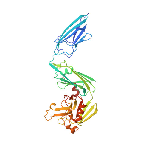 | |
UniProt | |||||
Entity Groups | |||||
| Sequence Clusters | 30% Identity50% Identity70% Identity90% Identity95% Identity100% Identity | ||||
| UniProt Group | A5U5L6 | ||||
Sequence AnnotationsExpand | |||||
Reference Sequence | |||||
| Ligands 4 Unique | |||||
|---|---|---|---|---|---|
| ID | Chains | Name / Formula / InChI Key | 2D Diagram | 3D Interactions | |
| E0Y Download:Ideal Coordinates CCD File | C [auth A], N [auth B] | (3S,5R)-5-[(2R,3R)-1,3-dihydroxybutan-2-yl]-3-({(3R)-1-[(1E)-ethanimidoyl]pyrrolidin-3-yl}sulfanyl)-L-proline C15 H27 N3 O4 S BSDSVSWWQWOWPI-SXFXLBHASA-N |  | ||
| PEG Download:Ideal Coordinates CCD File | D [auth A], O [auth B], P [auth B] | DI(HYDROXYETHYL)ETHER C4 H10 O3 MTHSVFCYNBDYFN-UHFFFAOYSA-N |  | ||
| SO4 Download:Ideal Coordinates CCD File | J [auth A], K [auth A], L [auth A], M [auth A], S [auth B] | SULFATE ION O4 S QAOWNCQODCNURD-UHFFFAOYSA-L |  | ||
| GOL Download:Ideal Coordinates CCD File | E [auth A] F [auth A] G [auth A] H [auth A] I [auth A] | GLYCEROL C3 H8 O3 PEDCQBHIVMGVHV-UHFFFAOYSA-N |  | ||
| Length ( Å ) | Angle ( ˚ ) |
|---|---|
| a = 61.06 | α = 90 |
| b = 94.06 | β = 92.7 |
| c = 75.4 | γ = 90 |
| Software Name | Purpose |
|---|---|
| PHENIX | refinement |
| DENZO | data reduction |
| SCALEPACK | data scaling |
| PHENIX | phasing |


