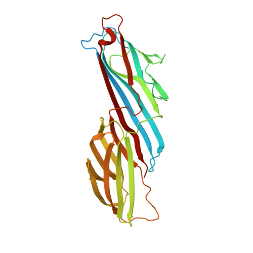
6BNT

Crystal structure of AP2 mu1 adaptin C-terminal domain with IRS-1 peptide


Experimental Data Snapshot

  • Method: X-RAY DIFFRACTION
  • Resolution: 3.20 Å
  • R-Value Free: 0.234 
  • R-Value Work: 0.206 
  • R-Value Observed: 0.209 

Starting Model: experimental
View more details

wwPDB Validation   3D Report Full Report


This is version 1.3 of the entry. See complete history


Literature

Mitotic regulators and the SHP2-MAPK pathway promote IR endocytosis and feedback regulation of insulin signaling.

Choi, E.Kikuchi, S.Gao, H.Brodzik, K.Nassour, I.Yopp, A.Singal, A.G.Zhu, H.Yu, H.

(2019) Nat Commun 10: 1473-1473

  • DOI: https://doi.org/10.1038/s41467-019-09318-3
  • Primary Citation of Related Structures:  
    6BNT

  • PubMed Abstract: 

    Insulin controls glucose homeostasis and cell growth through bifurcated signaling pathways. Dysregulation of insulin signaling is linked to diabetes and cancer. The spindle checkpoint controls the fidelity of chromosome segregation during mitosis. Here, we show that insulin receptor substrate 1 and 2 (IRS1/2) cooperate with spindle checkpoint proteins to promote insulin receptor (IR) endocytosis through recruiting the clathrin adaptor complex AP2 to IR. A phosphorylation switch of IRS1/2 orchestrated by extracellular signal-regulated kinase 1 and 2 (ERK1/2) and Src homology phosphatase 2 (SHP2) ensures selective internalization of activated IR. SHP2 inhibition blocks this feedback regulation and growth-promoting IR signaling, prolongs insulin action on metabolism, and improves insulin sensitivity in mice. We propose that mitotic regulators and SHP2 promote feedback inhibition of IR, thereby limiting the duration of insulin signaling. Targeting this feedback inhibition can improve insulin sensitivity.


  • Organizational Affiliation

    Howard Hughes Medical Institute, Department of Pharmacology, University of Texas Southwestern Medical Center, 6001 Forest Park Road, Dallas, TX, 75390, USA.


Macromolecules
Find similar proteins by:  (by identity cutoff)  |  3D Structure
Entity ID: 1
MoleculeChains Sequence LengthOrganismDetailsImage
AP-2 complex subunit mu315Homo sapiensMutation(s): 0 
Gene Names: AP2M1CLAPM1KIAA0109
UniProt & NIH Common Fund Data Resources
Find proteins for Q96CW1 (Homo sapiens)
Explore Q96CW1 
Go to UniProtKB:  Q96CW1
PHAROS:  Q96CW1
GTEx:  ENSG00000161203 
Entity Groups  
Sequence Clusters30% Identity50% Identity70% Identity90% Identity95% Identity100% Identity
UniProt GroupQ96CW1
Sequence Annotations
Expand
  • Reference Sequence

Find similar proteins by:  Sequence   |   3D Structure  

Entity ID: 2
MoleculeChains Sequence LengthOrganismDetailsImage
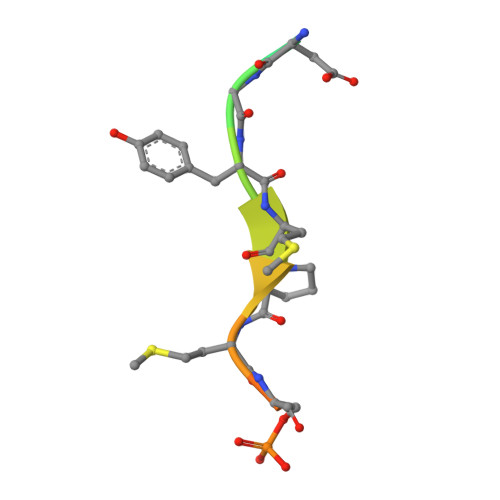
Insulin receptor substrate 115Homo sapiensMutation(s): 0 
Gene Names: IRS1
UniProt & NIH Common Fund Data Resources
Find proteins for P35568 (Homo sapiens)
Explore P35568 
Go to UniProtKB:  P35568
PHAROS:  P35568
GTEx:  ENSG00000169047 
Entity Groups  
Sequence Clusters30% Identity50% Identity70% Identity90% Identity95% Identity100% Identity
UniProt GroupP35568
Sequence Annotations
Expand
  • Reference Sequence
Small Molecules
Modified Residues  1 Unique
IDChains TypeFormula2D DiagramParent
SEP
Query on SEP
B
L-PEPTIDE LINKINGC3 H8 N O6 PSER
Experimental Data & Validation

Experimental Data

  • Method: X-RAY DIFFRACTION
  • Resolution: 3.20 Å
  • R-Value Free: 0.234 
  • R-Value Work: 0.206 
  • R-Value Observed: 0.209 
  • Space Group: P 64
Unit Cell:
Length ( Å )Angle ( ˚ )
a = 125.334α = 90
b = 125.334β = 90
c = 74.818γ = 120
Software Package:
Software NamePurpose
PHENIXrefinement
HKL-3000data reduction
HKL-3000data scaling
PHENIXphasing

Structure Validation

View Full Validation Report


Entry History & Funding Information

Deposition Data


Funding OrganizationLocationGrant Number
Clayton FoundationUnited States--

Revision History  (Full details and data files)

  • Version 1.0: 2018-11-21
    Type: Initial release
  • Version 1.1: 2019-06-05
    Changes: Data collection, Database references
  • Version 1.2: 2023-10-04
    Changes: Data collection, Database references, Refinement description
  • Version 1.3: 2024-10-30
    Changes: Structure summary