


Structure of Recombinant Dwarf Sperm Whale Myoglobin (Oxy)
Samuel, P.P., Miller, M.D., Xu, W., Alvarado, S., Phillips Jr., G.N., Olson, J.S.To be published.
Experimental Data Snapshot
Starting Model: experimental
View more details
Entity ID: 1 | |||||
|---|---|---|---|---|---|
| Molecule | Chains | Sequence Length | Organism | Details | Image |
| Myoglobin | 154 | Kogia sima | Mutation(s): 0 Gene Names: MB EC: 1.11.1 (UniProt), 1.7 (UniProt) | 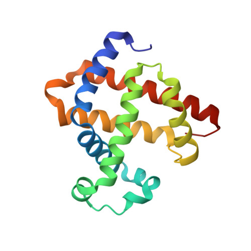 | |
UniProt | |||||
Entity Groups | |||||
| Sequence Clusters | 30% Identity50% Identity70% Identity90% Identity95% Identity100% Identity | ||||
| UniProt Group | P02184 | ||||
Sequence AnnotationsExpand | |||||
Reference Sequence | |||||
| Ligands 4 Unique | |||||
|---|---|---|---|---|---|
| ID | Chains | Name / Formula / InChI Key | 2D Diagram | 3D Interactions | |
| HEM Download:Ideal Coordinates CCD File | C [auth A], Q [auth B] | PROTOPORPHYRIN IX CONTAINING FE C34 H32 Fe N4 O4 KABFMIBPWCXCRK-RGGAHWMASA-L |  | ||
| CD Download:Ideal Coordinates CCD File | E [auth A] F [auth A] G [auth A] H [auth A] I [auth A] | CADMIUM ION Cd WLZRMCYVCSSEQC-UHFFFAOYSA-N |  | ||
| ACT Download:Ideal Coordinates CCD File | K [auth A] L [auth A] M [auth A] N [auth A] O [auth A] | ACETATE ION C2 H3 O2 QTBSBXVTEAMEQO-UHFFFAOYSA-M |  | ||
| OXY Download:Ideal Coordinates CCD File | D [auth A], R [auth B] | OXYGEN MOLECULE O2 MYMOFIZGZYHOMD-UHFFFAOYSA-N |  | ||
| Length ( Å ) | Angle ( ˚ ) |
|---|---|
| a = 85.995 | α = 90 |
| b = 85.995 | β = 90 |
| c = 109.01 | γ = 90 |
| Software Name | Purpose |
|---|---|
| PHENIX | refinement |
| XDS | data reduction |
| XDS | data scaling |
| Aimless | data scaling |
| PHASER | phasing |
| PDB_EXTRACT | data extraction |
| Funding Organization | Location | Grant Number |
|---|---|---|
| Robert A. Welch Foundation | United States | C-0612 |
| National Institutes of Health/National Institute of General Medical Sciences (NIH/NIGMS) | United States | R01-GM115261 |


