


Interaction of the rationally designed ritonavir-like inhibitors with human cytochrome P450 3A4: Impact of the side group interplay
Samuels, E.R., Sevrioukova, I.F.(2017) Mol Pharm
Experimental Data Snapshot
Starting Model: experimental
View more details
(2017) Mol Pharm
Entity ID: 1 | |||||
|---|---|---|---|---|---|
| Molecule | Chains | Sequence Length | Organism | Details | Image |
| Cytochrome P450 3A4 | 487 | Homo sapiens | Mutation(s): 0 Gene Names: CYP3A4, CYP3A3 EC: 1.14.13 (PDB Primary Data), 1.14.13.157 (PDB Primary Data), 1.14.13.32 (PDB Primary Data), 1.14.14.1 (PDB Primary Data), 1.14.13.67 (PDB Primary Data), 1.14.13.97 (PDB Primary Data), 1.14.14.73 (UniProt), 1.14.14.56 (UniProt), 1.14.14.55 (UniProt) | 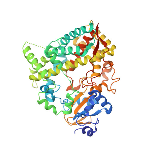 | |
UniProt & NIH Common Fund Data Resources | |||||
PHAROS: P08684 GTEx: ENSG00000160868 | |||||
Entity Groups | |||||
| Sequence Clusters | 30% Identity50% Identity70% Identity90% Identity95% Identity100% Identity | ||||
| UniProt Group | P08684 | ||||
Sequence AnnotationsExpand | |||||
Reference Sequence | |||||
| Ligands 2 Unique | |||||
|---|---|---|---|---|---|
| ID | Chains | Name / Formula / InChI Key | 2D Diagram | 3D Interactions | |
| HEM Download:Ideal Coordinates CCD File | B [auth A] | PROTOPORPHYRIN IX CONTAINING FE C34 H32 Fe N4 O4 KABFMIBPWCXCRK-RGGAHWMASA-L |  | ||
| Y83 Download:Ideal Coordinates CCD File | C [auth A] | tert-butyl [(2S)-1-(1H-indol-3-yl)-3-{[(2S)-3-oxo-2-(phenylamino)-3-{[(pyridin-3-yl)methyl]amino}propyl]sulfanyl}propan-2-yl]carbamate C31 H37 N5 O3 S YCHZAYWZVPKZNW-LBNVMWSVSA-N |  | ||
| Length ( Å ) | Angle ( ˚ ) |
|---|---|
| a = 77.29 | α = 90 |
| b = 101.41 | β = 90 |
| c = 127.37 | γ = 90 |
| Software Name | Purpose |
|---|---|
| PHENIX | refinement |
| MOSFLM | data reduction |
| SCALA | data scaling |
| PHASER | phasing |
| Funding Organization | Location | Grant Number |
|---|---|---|
| National Institutes of Health/National Institute of Environmental Health Sciences (NIH/NIEHS) | United States | ES025767 |


