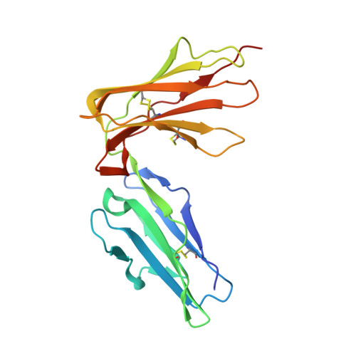
6BCS

LilrB2 D1D2 domains complexed with benzamidine


Experimental Data Snapshot

  • Method: X-RAY DIFFRACTION
  • Resolution: 2.10 Å
  • R-Value Free: 0.233 
  • R-Value Work: 0.182 
  • R-Value Observed: 0.187 

Starting Model: experimental
View more details

wwPDB Validation   3D Report Full Report


This is version 1.6 of the entry. See complete history


Literature

Inhibiting amyloid-beta cytotoxicity through its interaction with the cell surface receptor LilrB2 by structure-based design.

Cao, Q.Shin, W.S.Chan, H.Vuong, C.K.Dubois, B.Li, B.Murray, K.A.Sawaya, M.R.Feigon, J.Black, D.L.Eisenberg, D.S.Jiang, L.

(2018) Nat Chem 10: 1213-1221

  • DOI: https://doi.org/10.1038/s41557-018-0147-z
  • Primary Citation of Related Structures:  
    6BCS

  • PubMed Abstract: 

    Inhibiting the interaction between amyloid-β (Aβ) and a neuronal cell surface receptor, LilrB2, has been suggested as a potential route for treating Alzheimer's disease. Supporting this approach, Alzheimer's-like symptoms are reduced in mouse models following genetic depletion of the LilrB2 homologue. In its pathogenic, oligomeric state, Aβ binds to LilrB2, triggering a pathway to synaptic loss. Here we identify the LilrB2 binding moieties of Aβ ( 16 KLVFFA 21 ) and identify its binding site on LilrB2 from a crystal structure of LilrB2 immunoglobulin domains D1D2 complexed to small molecules that mimic phenylalanine residues. In this structure, we observed two pockets that can accommodate the phenylalanine side chains of KLVFFA. These pockets were confirmed to be 16 KLVFFA 21 binding sites by mutagenesis. Rosetta docking revealed a plausible geometry for the Aβ-LilrB2 complex and assisted with the structure-guided selection of small molecule inhibitors. These molecules inhibit Aβ-LilrB2 interactions in vitro and on the cell surface and reduce Aβ cytotoxicity, which suggests these inhibitors are potential therapeutic leads against Alzheimer's disease.


  • Organizational Affiliation

    Departments of Chemistry and Biochemistry and Biological Chemistry, UCLA-DOE Institute and Howard Hughes Medical Institute, UCLA, Los Angeles, CA, USA.


Macromolecules
Find similar proteins by:  (by identity cutoff)  |  3D Structure
Entity ID: 1
MoleculeChains Sequence LengthOrganismDetailsImage
Leukocyte immunoglobulin-like receptor subfamily B member 2198Homo sapiensMutation(s): 0 
Gene Names: LILRB2
UniProt & NIH Common Fund Data Resources
Find proteins for Q8N423 (Homo sapiens)
Explore Q8N423 
Go to UniProtKB:  Q8N423
PHAROS:  Q8N423
GTEx:  ENSG00000131042 
Entity Groups  
Sequence Clusters30% Identity50% Identity70% Identity90% Identity95% Identity100% Identity
UniProt GroupQ8N423
Sequence Annotations
Expand
  • Reference Sequence
Small Molecules
Ligands 3 Unique
IDChains Name / Formula / InChI Key2D Diagram3D Interactions
BEN
Query on BEN

Download Ideal Coordinates CCD File 
B [auth A],
C [auth A],
D [auth A],
E [auth A]
BENZAMIDINE
C7 H8 N2
PXXJHWLDUBFPOL-UHFFFAOYSA-N
DMS
Query on DMS

Download Ideal Coordinates CCD File 
H [auth A],
I [auth A]
DIMETHYL SULFOXIDE
C2 H6 O S
IAZDPXIOMUYVGZ-UHFFFAOYSA-N
CL
Query on CL

Download Ideal Coordinates CCD File 
F [auth A],
G [auth A]
CHLORIDE ION
Cl
VEXZGXHMUGYJMC-UHFFFAOYSA-M
Experimental Data & Validation

Experimental Data

  • Method: X-RAY DIFFRACTION
  • Resolution: 2.10 Å
  • R-Value Free: 0.233 
  • R-Value Work: 0.182 
  • R-Value Observed: 0.187 
  • Space Group: P 41 21 2
Unit Cell:
Length ( Å )Angle ( ˚ )
a = 57.05α = 90
b = 57.05β = 90
c = 101.18γ = 90
Software Package:
Software NamePurpose
XSCALEdata scaling
PHASERphasing
PHENIXrefinement
PDB_EXTRACTdata extraction
Cootmodel building
XDSdata reduction

Structure Validation

View Full Validation Report


Entry History & Funding Information

Deposition Data


Funding OrganizationLocationGrant Number
National Institutes of Health/National Institute on Aging (NIH/NIA)United StatesNIH AG054022

Revision History  (Full details and data files)

  • Version 1.0: 2018-09-05
    Type: Initial release
  • Version 1.1: 2018-10-24
    Changes: Data collection, Database references, Source and taxonomy
  • Version 1.2: 2018-12-05
    Changes: Data collection, Database references
  • Version 1.3: 2019-02-20
    Changes: Author supporting evidence, Data collection
  • Version 1.4: 2019-12-18
    Changes: Author supporting evidence
  • Version 1.5: 2023-10-04
    Changes: Data collection, Database references, Refinement description
  • Version 1.6: 2024-10-23
    Changes: Structure summary