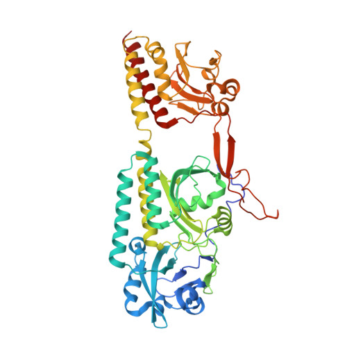
6BAP

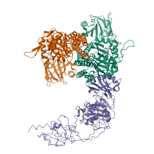
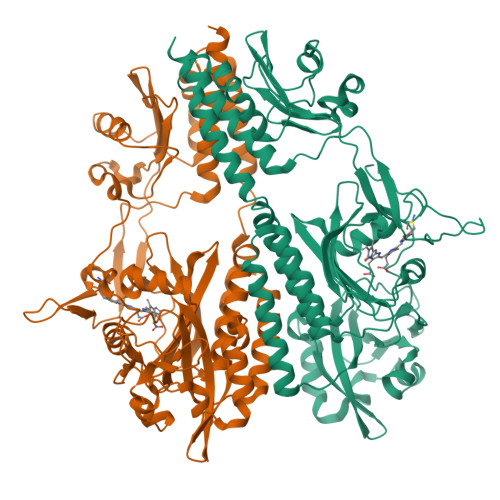
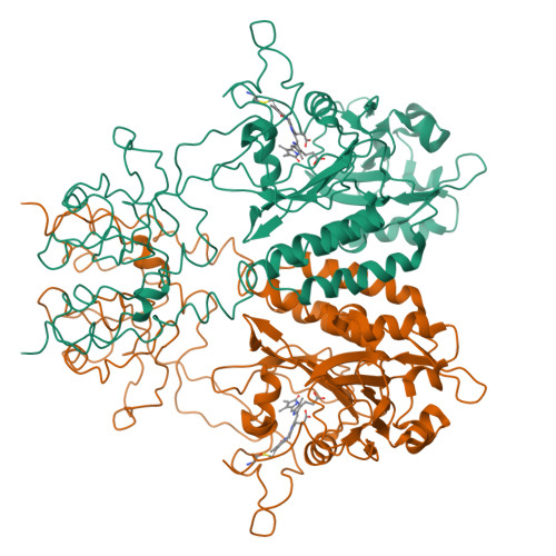
Stigmatella aurantiaca bacterial phytochrome PAS-GAF-PHY, T289H mutant

  • Classification: SIGNALING PROTEIN
  • Organism(s): Stigmatella aurantiaca DW4/3-1
  • Expression System: Escherichia coli
  • Mutation(s): Yes 

  • Deposited: 2017-10-14 Released: 2018-09-19 
  • Deposition Author(s): Schmidt, M., Stojkovic, E.
  • Funding Organization(s): National Science Foundation (NSF, United States), National Institutes of Health/National Institute of General Medical Sciences (NIH/NIGMS), National Institutes of Health/National Eye Institute (NIH/NEI)

Experimental Data Snapshot

  • Method: X-RAY DIFFRACTION
  • Resolution: 2.65 Å
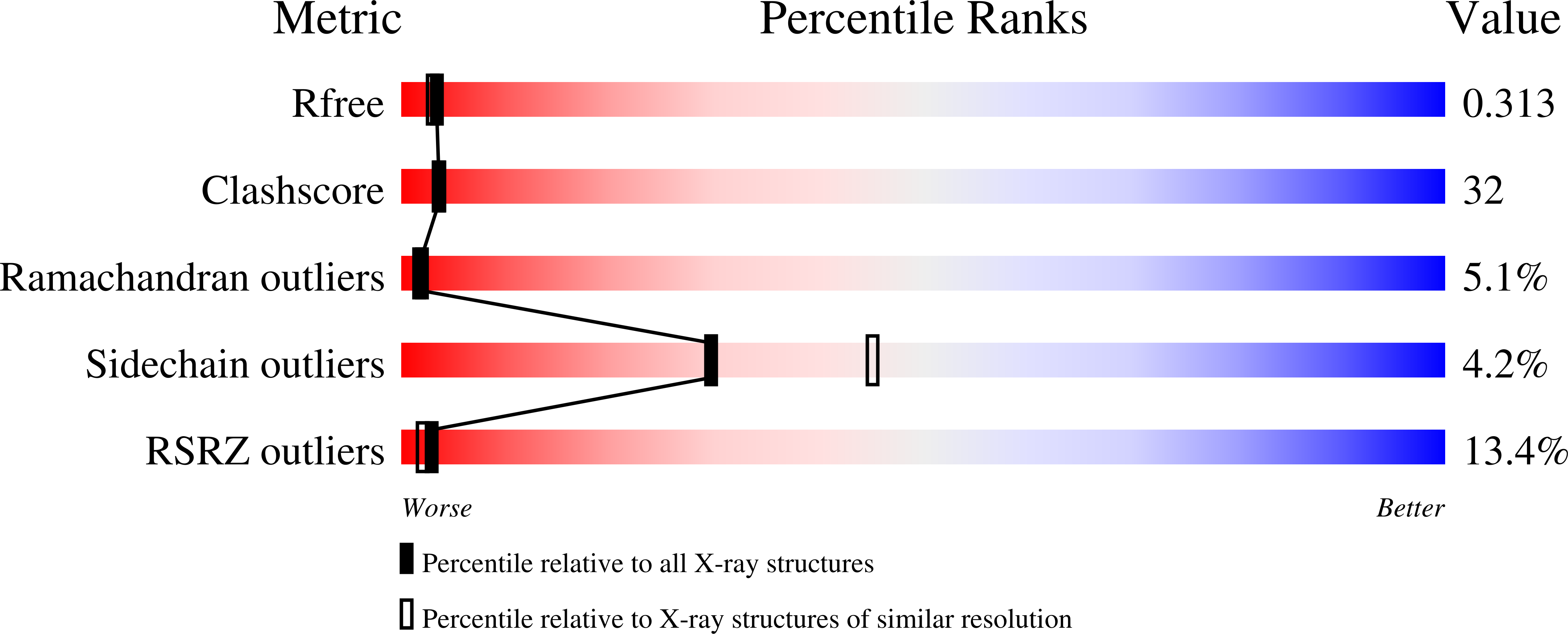
  • R-Value Free: 0.315 
  • R-Value Work: 0.241 
  • R-Value Observed: 0.245 

Starting Model: experimental
View more details

wwPDB Validation   3D Report Full Report


Ligand Structure Quality Assessment 


This is version 1.4 of the entry. See complete history


Literature

Structural basis for light control of cell development revealed by crystal structures of a myxobacterial phytochrome.

Woitowich, N.C.Halavaty, A.S.Waltz, P.Kupitz, C.Valera, J.Tracy, G.Gallagher, K.D.Claesson, E.Nakane, T.Pandey, S.Nelson, G.Tanaka, R.Nango, E.Mizohata, E.Owada, S.Tono, K.Joti, Y.Nugent, A.C.Patel, H.Mapara, A.Hopkins, J.Duong, P.Bizhga, D.Kovaleva, S.E.St Peter, R.Hernandez, C.N.Ozarowski, W.B.Roy-Chowdhuri, S.Yang, J.H.Edlund, P.Takala, H.Ihalainen, J.Brayshaw, J.Norwood, T.Poudyal, I.Fromme, P.Spence, J.C.H.Moffat, K.Westenhoff, S.Schmidt, M.Stojkovic, E.A.

(2018) IUCrJ 5: 619-634

  • DOI: https://doi.org/10.1107/S2052252518010631
  • Primary Citation of Related Structures:  
    6BAF, 6BAK, 6BAO, 6BAP, 6BAY

  • PubMed Abstract: 

    Phytochromes are red-light photoreceptors that were first characterized in plants, with homologs in photosynthetic and non-photosynthetic bacteria known as bacteriophytochromes (BphPs). Upon absorption of light, BphPs interconvert between two states denoted Pr and Pfr with distinct absorption spectra in the red and far-red. They have recently been engineered as enzymatic photoswitches for fluorescent-marker applications in non-invasive tissue imaging of mammals. This article presents cryo- and room-temperature crystal structures of the unusual phytochrome from the non-photosynthetic myxo-bacterium Stigmatella aurantiaca (SaBphP1) and reveals its role in the fruiting-body formation of this photomorphogenic bacterium. SaBphP1 lacks a conserved histidine (His) in the chromophore-binding domain that stabilizes the Pr state in the classical BphPs. Instead it contains a threonine (Thr), a feature that is restricted to several myxobacterial phytochromes and is not evolutionarily understood. SaBphP1 structures of the chromophore binding domain (CBD) and the complete photosensory core module (PCM) in wild-type and Thr-to-His mutant forms reveal details of the molecular mechanism of the Pr/Pfr transition associated with the physiological response of this myxobacterium to red light. Specifically, key structural differences in the CBD and PCM between the wild-type and the Thr-to-His mutant involve essential chromophore contacts with proximal amino acids, and point to how the photosignal is transduced through the rest of the protein, impacting the essential enzymatic activity in the photomorphogenic response of this myxobacterium.


  • Organizational Affiliation

    Department of Biology, Northeastern Illinois University, Chicago, IL, USA.


Macromolecules
Find similar proteins by:  (by identity cutoff)  |  3D Structure
Entity ID: 1
MoleculeChains Sequence LengthOrganismDetailsImage
Photoreceptor-histidine kinase BphP
A, B, C
515Stigmatella aurantiaca DW4/3-1Mutation(s): 1 
Gene Names: STAUR_8015STIAU_3396
EC: 2.7.13.3
UniProt
Find proteins for Q097N3 (Stigmatella aurantiaca (strain DW4/3-1))
Explore Q097N3 
Go to UniProtKB:  Q097N3
Entity Groups  
Sequence Clusters30% Identity50% Identity70% Identity90% Identity95% Identity100% Identity
UniProt GroupQ097N3
Sequence Annotations
Expand
  • Reference Sequence
Small Molecules
Ligands 1 Unique
IDChains Name / Formula / InChI Key2D Diagram3D Interactions
BLR
Query on BLR

Download Ideal Coordinates CCD File 
D [auth A],
E [auth B],
F [auth C]
3-[5-[(Z)-(4-ethenyl-3-methyl-5-oxidanylidene-pyrrol-2-ylidene)methyl]-2-[[5-[(Z)-(3-ethenyl-4-methyl-5-oxidanylidene-pyrrol-2-ylidene)methyl]-3-(3-hydroxy-3-oxopropyl)-4-methyl-1H-pyrrol-2-yl]methyl]-4-methyl-1H-pyrrol-3-yl]propanoic acid
C33 H36 N4 O6
BPYKTIZUTYGOLE-IFADSCNNSA-N
Experimental Data & Validation

Experimental Data

  • Method: X-RAY DIFFRACTION
  • Resolution: 2.65 Å
  • R-Value Free: 0.315 
  • R-Value Work: 0.241 
  • R-Value Observed: 0.245 
  • Space Group: P 32 2 1
Unit Cell:
Length ( Å )Angle ( ˚ )
a = 83.31α = 90
b = 83.31β = 90
c = 475.05γ = 120
Software Package:
Software NamePurpose
PHENIXrefinement
HKL-2000data reduction
MOSFLMdata reduction
SCALAdata scaling
PHASERphasing

Structure Validation

View Full Validation Report


Ligand Structure Quality Assessment 


Entry History & Funding Information

Deposition Data


Funding OrganizationLocationGrant Number
National Science Foundation (NSF, United States)United StatesSTC-1231306
National Science Foundation (NSF, United States)United StatesBIO-MCB 1413360
National Institutes of Health/National Institute of General Medical Sciences (NIH/NIGMS)United StatesT34 GM105549-01 NU-STARS
National Institutes of Health/National Eye Institute (NIH/NEI)United StatesEY024363

Revision History  (Full details and data files)

  • Version 1.0: 2018-09-19
    Type: Initial release
  • Version 1.1: 2018-10-31
    Changes: Data collection, Database references
  • Version 1.2: 2019-02-20
    Changes: Author supporting evidence, Data collection
  • Version 1.3: 2019-11-27
    Changes: Author supporting evidence
  • Version 1.4: 2023-10-04
    Changes: Data collection, Database references, Refinement description