6B7H | pdb_00006b7h

Structure of mGluR3 with an agonist


Experimental Data Snapshot

  • Method: X-RAY DIFFRACTION
  • Resolution: 2.82 Å
  • R-Value Free: 
    0.223 (Depositor), 0.247 (DCC) 
  • R-Value Work: 
    0.184 (Depositor), 0.203 (DCC) 
  • R-Value Observed: 
    0.185 (Depositor) 

wwPDB Validation 3D Report Full Report

Validation slider image for 6B7H

Ligand Structure Quality Assessment 


This is version 2.1 of the entry. See complete history

Literature

Synthesis and Pharmacological Characterization of C4beta-Amide-Substituted 2-Aminobicyclo[3.1.0]hexane-2,6-dicarboxylates. Identification of (1 S,2 S,4 S,5 R,6 S)-2-Amino-4-[(3-methoxybenzoyl)amino]bicyclo[3.1.0]hexane-2,6-dicarboxylic Acid (LY2794193), a Highly Potent and Selective mGlu3Receptor Agonist.

Monn, J.A.Henry, S.S.Massey, S.M.Clawson, D.K.Chen, Q.Diseroad, B.A.Bhardwaj, R.M.Atwell, S.Lu, F.Wang, J.Russell, M.Heinz, B.A.Wang, X.S.Carter, J.H.Getman, B.G.Adragni, K.Broad, L.M.Sanger, H.E.Ursu, D.Catlow, J.T.Swanson, S.Johnson, B.G.Shaw, D.B.McKinzie, D.L.Hao, J.

(2018) J Med Chem 61: 2303-2328

  • DOI: https://doi.org/10.1021/acs.jmedchem.7b01481
  • Primary Citation Related Structures: 
    6B7H

  • PubMed Abstract: 

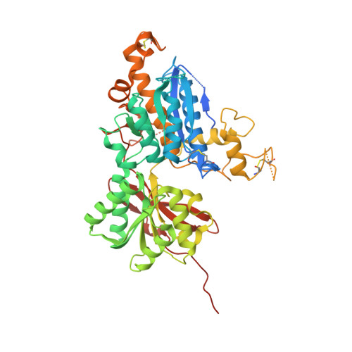
    Multiple therapeutic opportunities have been suggested for compounds capable of selective activation of metabotropic glutamate 3 (mGlu 3 ) receptors, but small molecule tools are lacking. As part of our ongoing efforts to identify potent, selective, and systemically bioavailable agonists for mGlu 2 and mGlu 3 receptor subtypes, a series of C4 β -N-linked variants of (1 S,2 S,5 R,6 S)-2-amino-bicyclo[3.1.0]hexane-2,6-dicarboxylic acid 1 (LY354740) were prepared and evaluated for both mGlu 2 and mGlu 3 receptor binding affinity and functional cellular responses. From this investigation we identified (1 S,2 S,4 S,5 R,6 S)-2-amino-4-[(3-methoxybenzoyl)amino]bicyclo[3.1.0]hexane-2,6-dicarboxylic acid 8p (LY2794193), a molecule that demonstrates remarkable mGlu 3 receptor selectivity. Crystallization of 8p with the amino terminal domain of hmGlu 3 revealed critical binding interactions for this ligand with residues adjacent to the glutamate binding site, while pharmacokinetic assessment of 8p combined with its effect in an mGlu 2 receptor-dependent behavioral model provides estimates for doses of this compound that would be expected to selectively engage and activate central mGlu 3 receptors in vivo.

Macromolecule Content 

  • Total Structure Weight: 60.22 kDa 
  • Atom Count: 3,567 
  • Modeled Residue Count: 441 
  • Deposited Residue Count: 517 
  • Unique protein chains: 1

Macromolecules

Find similar proteins by:|  3D Structure
Entity ID: 1
MoleculeChains  Sequence LengthOrganismDetailsImage
Metabotropic glutamate receptor 3517Homo sapiensMutation(s): 0 
Gene Names: GRM3GPRC1CMGLUR3
UniProt & NIH Common Fund Data Resources
Find proteins for Q14832 (Homo sapiens)
Explore Q14832 
Go to UniProtKB:  Q14832
PHAROS:  Q14832
GTEx:  ENSG00000198822 
Entity Groups
Sequence Clusters30% Identity50% Identity70% Identity90% Identity95% Identity100% Identity
UniProt GroupQ14832
Glycosylation
Glycosylation Sites: 3Go to GlyGen: Q14832-1
Sequence Annotations
Expand
Reference Sequence

Oligosaccharides

Help  
Entity ID: 2
MoleculeChains Length2D Diagram GlycosylationD Interactions
2-acetamido-2-deoxy-beta-D-glucopyranose-(1-4)-2-acetamido-2-deoxy-beta-D-glucopyranose
B, C
2N-Glycosylation
Glycosylation Resources
GlyTouCan: G42666HT
GlyCosmos: G42666HT
GlyGen: G42666HT

Small Molecules

Ligands 3 Unique
IDChains Name / Formula / InChI Key2D Diagram3D Interactions
CWY

Query on CWY


Download:Ideal Coordinates CCD File
E [auth A](1S,2S,4S,5R,6S)-2-amino-4-[(3-methoxybenzene-1-carbonyl)amino]bicyclo[3.1.0]hexane-2,6-dicarboxylic acid
C16 H18 N2 O6
UXNRHIJPZNNDDJ-VZAVHYRXSA-N
NAG

Query on NAG


Download:Ideal Coordinates CCD File
D [auth A]2-acetamido-2-deoxy-beta-D-glucopyranose
C8 H15 N O6
OVRNDRQMDRJTHS-FMDGEEDCSA-N
CA

Query on CA


Download:Ideal Coordinates CCD File
F [auth A]CALCIUM ION
Ca
BHPQYMZQTOCNFJ-UHFFFAOYSA-N
Binding Affinity Annotations 
IDSourceBinding Affinity
CWY BindingDB:  6B7H Ki: min: 0.93, max: 106 (nM) from 2 assay(s)
EC50: min: 0.47, max: 12 (nM) from 2 assay(s)

Experimental Data & Validation

Experimental Data

  • Method: X-RAY DIFFRACTION
  • Resolution: 2.82 Å
  • R-Value Free:  0.223 (Depositor), 0.247 (DCC) 
  • R-Value Work:  0.184 (Depositor), 0.203 (DCC) 
  • R-Value Observed: 0.185 (Depositor) 
Space Group: P 32 2 1
Unit Cell:
Length ( Å )Angle ( ˚ )
a = 80.081α = 90
b = 80.081β = 90
c = 161.887γ = 120
Software Package:
Software NamePurpose
MOSFLMdata collection
SCALAdata scaling
BUSTERrefinement
PDB_EXTRACTdata extraction
SCALAdata reduction
PHASERphasing

Structure Validation

View Full Validation Report


Ligand Structure Quality Assessment 


Entry History 

Deposition Data

Revision History  (Full details and data files)

  • Version 1.0: 2018-04-25
    Type: Initial release
  • Version 2.0: 2020-07-29
    Type: Remediation
    Reason: Carbohydrate remediation
    Changes: Atomic model, Data collection, Derived calculations, Structure summary
  • Version 2.1: 2024-10-23
    Changes: Data collection, Database references, Structure summary