A comparative study of the binding properties, dipeptidyl peptidase-4 (DPP-4) inhibitory activity and glucose-lowering efficacy of the DPP-4 inhibitors alogliptin, linagliptin, saxagliptin, sitagliptin and vildagliptin in mice.
Berger, J.P., SinhaRoy, R., Pocai, A., Kelly, T.M., Scapin, G., Gao, Y.D., Pryor, K.A.D., Wu, J.K., Eiermann, G.J., Xu, S.S., Zhang, X., Tatosian, D.A., Weber, A.E., Thornberry, N.A., Carr, R.D.(2018) Endocrinol Diabetes Metab 1: e00002-e00002
- PubMed: 30815539 Search on PubMedSearch on PubMed Central
- DOI: https://doi.org/10.1002/edm2.2
- Primary Citation Related Structures:
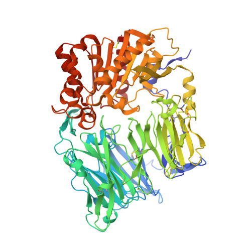
6B1E, 6B1O - PubMed Abstract:
Since 2006, DPP-4 inhibitors have become established therapy for the treatment of type 2 diabetes. Despite sharing a common mechanism of action, considerable chemical diversity exists amongst members of the DPP-4 inhibitor class, raising the question as to whether structural differences may result in differentiated enzyme inhibition and antihyperglycaemic activity. We have compared the binding properties of the most commonly used inhibitors and have investigated the relationship between their inhibitory potency at the level of the enzyme and their acute glucose-lowering efficacy. Firstly, using a combination of published crystal structures and in-house data, we demonstrated that the binding site utilized by all of the DPP-4 inhibitors assessed was the same as that used by neuropeptide Y, supporting the hypothesis that DPP-4 inhibitors are able to competitively inhibit endogenous substrates for the enzyme. Secondly, we ascertained that the enzymatic cleft of DPP-4 is a relatively large cavity which displays conformational flexibility to accommodate structurally diverse inhibitor molecules. Finally, we found that for all inhibitors, irrespective of their chemical structure, the inhibition of plasma DPP-4 enzyme activity correlates directly with acute plasma glucose lowering in mice. The common binding site utilized by different DPP-4 inhibitors enables similar competitive inhibition of the cleavage of the endogenous DPP-4 substrates. Furthermore, despite chemical diversity and a range of binding potencies observed amongst the DPP-4 inhibitors, a direct relationship between enzyme inhibition in the plasma and glucose lowering is evident in mice for each member of the classes studied.
- Merck& Co., Inc. Kenilworth NJ USA.
Organizational Affiliation: