6B0X | pdb_00006b0x

Capsid protein and C-terminal part of scaffolding protein in the Staphylococcus aureus phage 80alpha procapsid


Experimental Data Snapshot

  • Method: ELECTRON MICROSCOPY
  • Resolution: 3.80 Å
  • Aggregation State: PARTICLE 
  • Reconstruction Method: SINGLE PARTICLE 

wwPDB Validation 3D Report Full Report

Validation slider image for 6B0X

This is version 1.4 of the entry. See complete history

Literature

Competing scaffolding proteins determine capsid size during mobilization ofStaphylococcus aureuspathogenicity islands.

Dearborn, A.D.Wall, E.A.Kizziah, J.L.Klenow, L.Parker, L.K.Manning, K.A.Spilman, M.S.Spear, J.M.Christie, G.E.Dokland, T.

(2017) Elife 6

  • DOI: https://doi.org/10.7554/eLife.30822
  • Primary Citation Related Structures: 
    6B0X, 6B23

  • PubMed Abstract: 

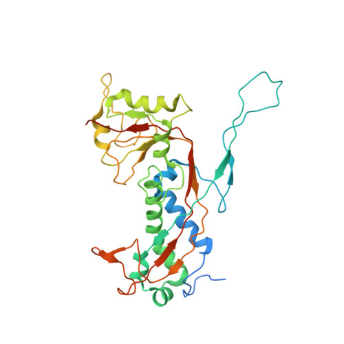
    Staphylococcus aureus pathogenicity islands (SaPIs), such as SaPI1, exploit specific helper bacteriophages, like 80α, for their high frequency mobilization, a process termed 'molecular piracy'. SaPI1 redirects the helper's assembly pathway to form small capsids that can only accommodate the smaller SaPI1 genome, but not a complete phage genome. SaPI1 encodes two proteins, CpmA and CpmB, that are responsible for this size redirection. We have determined the structures of the 80α and SaPI1 procapsids to near-atomic resolution by cryo-electron microscopy, and show that CpmB competes with the 80α scaffolding protein (SP) for a binding site on the capsid protein (CP), and works by altering the angle between capsomers. We probed these interactions genetically and identified second-site suppressors of lethal mutations in SP. Our structures show, for the first time, the detailed interactions between SP and CP in a bacteriophage, providing unique insights into macromolecular assembly processes.


  • Organizational Affiliation
    • Protein Expression Laboratory, National Institute of Arthritis and Musculoskeletal and Skin Diseases, National Institutes of Health, Bethesda, United States.

Macromolecule Content 

  • Total Structure Weight: 421.8 kDa 
  • Atom Count: 16,791 
  • Modeled Residue Count: 2,097 
  • Deposited Residue Count: 3,710 
  • Unique protein chains: 2

Macromolecules

Find similar proteins by:|  3D Structure
Entity ID: 1
MoleculeChains  Sequence LengthOrganismDetailsImage
Major head protein
A, B, C, D, E
A, B, C, D, E, F, G
324Dubowvirus dv80alphaMutation(s): 0 
Gene Names: orf47
UniProt
Find proteins for A4ZFB3 (Staphylococcus phage 80alpha)
Explore A4ZFB3 
Go to UniProtKB:  A4ZFB3
Entity Groups
Sequence Clusters30% Identity50% Identity70% Identity90% Identity95% Identity100% Identity
UniProt GroupA4ZFB3
Sequence Annotations
Expand
Reference Sequence
Find similar proteins by:|  3D Structure
Entity ID: 2
MoleculeChains  Sequence LengthOrganismDetailsImage
Scaffold protein206Dubowvirus dv80alphaMutation(s): 0 
UniProt
Find proteins for A4ZFB2 (Staphylococcus phage 80alpha)
Explore A4ZFB2 
Go to UniProtKB:  A4ZFB2
Entity Groups
Sequence Clusters30% Identity50% Identity70% Identity90% Identity95% Identity100% Identity
UniProt GroupA4ZFB2
Sequence Annotations
Expand
Reference Sequence

Experimental Data & Validation

Experimental Data

  • Method: ELECTRON MICROSCOPY
  • Resolution: 3.80 Å
  • Aggregation State: PARTICLE 
  • Reconstruction Method: SINGLE PARTICLE 

Structure Validation

View Full Validation Report


Entry History 

& Funding Information

Deposition Data


Funding OrganizationLocationGrant Number
National Institutes of Health/National Institute Of Allergy and Infectious Diseases (NIH/NIAID)United StatesR01AI083255

Revision History  (Full details and data files)

  • Version 1.0: 2017-10-18
    Type: Initial release
  • Version 1.1: 2018-02-28
    Changes: Database references, Structure summary
  • Version 1.2: 2018-03-28
    Changes: Data collection, Database references
  • Version 1.3: 2019-12-11
    Changes: Author supporting evidence
  • Version 1.4: 2024-03-13
    Changes: Data collection, Database references, Derived calculations, Refinement description