6ATT | pdb_00006att

39S Fab bound to HER2 ecd


Experimental Data Snapshot

  • Method: X-RAY DIFFRACTION
  • Resolution: 3.77 Å
  • R-Value Free: 
    0.289 (Depositor), 0.293 (DCC) 
  • R-Value Work: 
    0.251 (Depositor), 0.265 (DCC) 
  • R-Value Observed: 
    0.253 (Depositor) 

Starting Model: experimental
View more details

wwPDB Validation 3D Report Full Report

Validation slider image for 6ATT

Ligand Structure Quality Assessment 


This is version 2.2 of the entry. See complete history

Literature

Structural insights into the mechanism of action of a biparatopic anti-HER2 antibody.

Oganesyan, V.Peng, L.Bee, J.S.Li, J.Perry, S.R.Comer, F.Xu, L.Cook, K.Senthil, K.Clarke, L.Rosenthal, K.Gao, C.Damschroder, M.Wu, H.Dall'Acqua, W.

(2018) J Biological Chem 293: 8439-8448

  • DOI: https://doi.org/10.1074/jbc.M117.818013
  • Primary Citation Related Structures: 
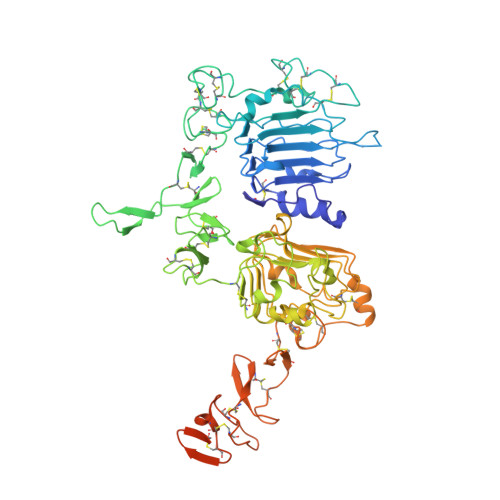
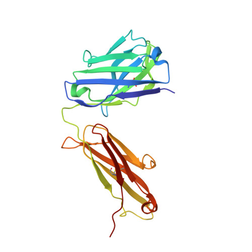
    6ATT

  • PubMed Abstract: 

    Pathways of human epidermal growth factor (EGF) receptors are activated upon ligand-dependent or -independent homo- or heterodimerization and their subsequent transphosphorylation. Overexpression of these receptors positively correlates with transphosphorylation rates and increased tumor growth rates. MEDI4276, an anti-human epidermal growth factor receptor 2 (HER2) biparatopic antibody-drug conjugate, has two paratopes within each antibody arm. One, 39S, is aiming at the HER2 site involved in receptor dimerization and the second, single chain fragment (scFv), mimicking trastuzumab. Here we present the cocrystal structure of the 39S Fab-HER2 complex and, along with biophysical and functional assays, determine the corresponding epitope of MEDI4276 and its underlying mechanism of action. Our results reveal that MEDI4276's uniqueness is based first on the ability of its 39S paratope to block HER2 homo- or heterodimerization and second on its ability to cluster the receptors on the surface of receptor-overexpressing cells.


  • Organizational Affiliation
    • From the Departments of Antibody Discovery and Protein Engineering, oganesyanv@medimmune.com.

Macromolecule Content 

  • Total Structure Weight: 118.52 kDa 
  • Atom Count: 7,819 
  • Modeled Residue Count: 1,011 
  • Deposited Residue Count: 1,074 
  • Unique protein chains: 3

Macromolecules

Find similar proteins by:|  3D Structure
Entity ID: 1
MoleculeChains  Sequence LengthOrganismDetailsImage
Receptor tyrosine-protein kinase erbB-2630Homo sapiensMutation(s): 0 
Gene Names: ERBB2HER2MLN19NEUNGL
EC: 2.7.10.1
UniProt & NIH Common Fund Data Resources
Find proteins for P04626 (Homo sapiens)
Explore P04626 
Go to UniProtKB:  P04626
PHAROS:  P04626
GTEx:  ENSG00000141736 
Entity Groups
Sequence Clusters30% Identity50% Identity70% Identity90% Identity95% Identity100% Identity
UniProt GroupP04626
Glycosylation
Glycosylation Sites: 3Go to GlyGen: P04626-1
Sequence Annotations
Expand
Reference Sequence
Find similar proteins by:|  3D Structure
Entity ID: 2
MoleculeChains  Sequence LengthOrganismDetailsImage
Antibody 39S Fab heavy chainB [auth H]224Homo sapiensMutation(s): 0 
Find similar proteins by:|  3D Structure
Entity ID: 3
MoleculeChains  Sequence LengthOrganismDetailsImage
Antibody 39S Fab light chainC [auth L]220Homo sapiensMutation(s): 0 

Oligosaccharides

Help  
Entity ID: 4
MoleculeChains Length2D Diagram GlycosylationD Interactions
2-acetamido-2-deoxy-beta-D-glucopyranose-(1-4)-2-acetamido-2-deoxy-beta-D-glucopyranoseD [auth B],
E [auth C]
2N-Glycosylation
Glycosylation Resources
GlyTouCan: G42666HT
GlyCosmos: G42666HT
GlyGen: G42666HT

Small Molecules

Ligands 1 Unique
IDChains Name / Formula / InChI Key2D Diagram3D Interactions
NAG

Query on NAG


Download:Ideal Coordinates CCD File
F [auth A]2-acetamido-2-deoxy-beta-D-glucopyranose
C8 H15 N O6
OVRNDRQMDRJTHS-FMDGEEDCSA-N

Experimental Data & Validation

Experimental Data

  • Method: X-RAY DIFFRACTION
  • Resolution: 3.77 Å
  • R-Value Free:  0.289 (Depositor), 0.293 (DCC) 
  • R-Value Work:  0.251 (Depositor), 0.265 (DCC) 
  • R-Value Observed: 0.253 (Depositor) 
Space Group: P 21 21 21
Unit Cell:
Length ( Å )Angle ( ˚ )
a = 67.327α = 90
b = 108.172β = 90
c = 348.308γ = 90
Software Package:
Software NamePurpose
REFMACrefinement
HKL-2000data reduction
SCALEPACKdata scaling
MOLREPphasing

Structure Validation

View Full Validation Report


Ligand Structure Quality Assessment 


Entry History 

Deposition Data

Revision History  (Full details and data files)

  • Version 1.0: 2018-04-25
    Type: Initial release
  • Version 1.1: 2018-05-02
    Changes: Data collection, Database references
  • Version 1.2: 2018-06-13
    Changes: Data collection, Database references
  • Version 2.0: 2020-07-29
    Type: Remediation
    Reason: Carbohydrate remediation
    Changes: Advisory, Atomic model, Data collection, Derived calculations, Structure summary
  • Version 2.1: 2023-10-04
    Changes: Data collection, Database references, Refinement description, Structure summary
  • Version 2.2: 2024-10-09
    Changes: Structure summary