6AQF | pdb_00006aqf

Crystal structure of A2AAR-BRIL in complex with the antagonist ZM241385 produced from Pichia pastoris


Experimental Data Snapshot

  • Method: X-RAY DIFFRACTION
  • Resolution: 2.51 Å
  • R-Value Free: 
    0.256 (Depositor), 0.272 (DCC) 
  • R-Value Work: 
    0.220 (Depositor), 0.231 (DCC) 
  • R-Value Observed: 
    0.222 (Depositor) 

Starting Model: experimental
View more details

wwPDB Validation 3D Report Full Report

Validation slider image for 6AQF

Ligand Structure Quality Assessment 


This is version 1.4 of the entry. See complete history

Literature

Allosteric Coupling of Drug Binding and Intracellular Signaling in the A2A Adenosine Receptor.

Eddy, M.T.Lee, M.Y.Gao, Z.G.White, K.L.Didenko, T.Horst, R.Audet, M.Stanczak, P.McClary, K.M.Han, G.W.Jacobson, K.A.Stevens, R.C.Wuthrich, K.

(2018) Cell 172: 68-80.e12

  • DOI: https://doi.org/10.1016/j.cell.2017.12.004
  • Primary Citation Related Structures: 
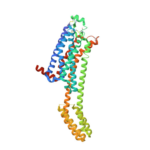
    6AQF

  • PubMed Abstract: 

    Signaling across cellular membranes, the 826 human G protein-coupled receptors (GPCRs) govern a wide range of vital physiological processes, making GPCRs prominent drug targets. X-ray crystallography provided GPCR molecular architectures, which also revealed the need for additional structural dynamics data to support drug development. Here, nuclear magnetic resonance (NMR) spectroscopy with the wild-type-like A 2A adenosine receptor (A 2A AR) in solution provides a comprehensive characterization of signaling-related structural dynamics. All six tryptophan indole and eight glycine backbone 15 N- 1 H NMR signals in A 2A AR were individually assigned. These NMR probes provided insight into the role of Asp52 2.50 as an allosteric link between the orthosteric drug binding site and the intracellular signaling surface, revealing strong interactions with the toggle switch Trp 246 6.48 , and delineated the structural response to variable efficacy of bound drugs across A 2A AR. The present data support GPCR signaling based on dynamic interactions between two semi-independent subdomains connected by an allosteric switch at Asp52 2.50 .


  • Organizational Affiliation
    • Department of Integrative Structural and Computational Biology, The Scripps Research Institute, La Jolla, CA 92037, USA; Departments of Biological Sciences and Chemistry, Bridge Institute, University of Southern California, Los Angeles, CA 90089, USA.

Macromolecule Content 

  • Total Structure Weight: 53.55 kDa 
  • Atom Count: 3,263 
  • Modeled Residue Count: 390 
  • Deposited Residue Count: 432 
  • Unique protein chains: 1

Macromolecules

Find similar proteins by:|  3D Structure
Entity ID: 1
MoleculeChains  Sequence LengthOrganismDetailsImage
Adenosine receptor A2a,Soluble cytochrome b562,Adenosine receptor A2a432Homo sapiensEscherichia coliMutation(s): 3 
Gene Names: ADORA2AADORA2cybC
Membrane Entity: Yes 
UniProt & NIH Common Fund Data Resources
Find proteins for P0ABE7 (Escherichia coli)
Explore P0ABE7 
Go to UniProtKB:  P0ABE7
Find proteins for P29274 (Homo sapiens)
Explore P29274 
Go to UniProtKB:  P29274
PHAROS:  P29274
GTEx:  ENSG00000128271 
Entity Groups
Sequence Clusters30% Identity50% Identity70% Identity90% Identity95% Identity100% Identity
UniProt GroupsP0ABE7P29274
Sequence Annotations
Expand
Reference Sequence

Small Molecules

Ligands 5 Unique
IDChains Name / Formula / InChI Key2D Diagram3D Interactions
CLR

Query on CLR


Download:Ideal Coordinates CCD File
C [auth A],
D [auth A],
E [auth A]
CHOLESTEROL
C27 H46 O
HVYWMOMLDIMFJA-DPAQBDIFSA-N
OLC
(Subject of Investigation/LOI)

Query on OLC


Download:Ideal Coordinates CCD File
N [auth A],
O [auth A],
P [auth A],
Q [auth A]
(2R)-2,3-dihydroxypropyl (9Z)-octadec-9-enoate
C21 H40 O4
RZRNAYUHWVFMIP-GDCKJWNLSA-N
OLB
(Subject of Investigation/LOI)

Query on OLB


Download:Ideal Coordinates CCD File
F [auth A](2S)-2,3-dihydroxypropyl (9Z)-octadec-9-enoate
C21 H40 O4
RZRNAYUHWVFMIP-QJRAZLAKSA-N
ZMA
(Subject of Investigation/LOI)

Query on ZMA


Download:Ideal Coordinates CCD File
B [auth A]4-{2-[(7-amino-2-furan-2-yl[1,2,4]triazolo[1,5-a][1,3,5]triazin-5-yl)amino]ethyl}phenol
C16 H15 N7 O2
PWTBZOIUWZOPFT-UHFFFAOYSA-N
OLA
(Subject of Investigation/LOI)

Query on OLA


Download:Ideal Coordinates CCD File
G [auth A]
H [auth A]
I [auth A]
J [auth A]
K [auth A]
G [auth A],
H [auth A],
I [auth A],
J [auth A],
K [auth A],
L [auth A],
M [auth A]
OLEIC ACID
C18 H34 O2
ZQPPMHVWECSIRJ-KTKRTIGZSA-N

Experimental Data & Validation

Experimental Data

  • Method: X-RAY DIFFRACTION
  • Resolution: 2.51 Å
  • R-Value Free:  0.256 (Depositor), 0.272 (DCC) 
  • R-Value Work:  0.220 (Depositor), 0.231 (DCC) 
  • R-Value Observed: 0.222 (Depositor) 
Space Group: C 2 2 21
Unit Cell:
Length ( Å )Angle ( ˚ )
a = 39.837α = 90
b = 180.973β = 90
c = 140.574γ = 90
Software Package:
Software NamePurpose
BUSTERrefinement
HKL-2000data reduction
HKL-2000data scaling
PHASERphasing

Structure Validation

View Full Validation Report


Ligand Structure Quality Assessment 


Entry History 

Revision History  (Full details and data files)

  • Version 1.0: 2018-01-10
    Type: Initial release
  • Version 1.1: 2018-01-17
    Changes: Database references
  • Version 1.2: 2018-01-24
    Changes: Database references
  • Version 1.3: 2023-10-04
    Changes: Data collection, Database references, Refinement description
  • Version 1.4: 2024-10-16
    Changes: Structure summary