6APL | pdb_00006apl

Crystal Structure of human ST6GALNAC2 in complex with CMP


Experimental Data Snapshot

  • Method: X-RAY DIFFRACTION
  • Resolution: 2.35 Å
  • R-Value Free: 
    0.236 (Depositor), 0.238 (DCC) 
  • R-Value Work: 
    0.196 (Depositor), 0.199 (DCC) 
  • R-Value Observed: 
    0.200 (Depositor) 

Starting Model: experimental
View more details

wwPDB Validation 3D Report Full Report

Validation slider image for 6APL

Ligand Structure Quality Assessment 


This is version 1.5 of the entry. See complete history

Literature

Expression system for structural and functional studies of human glycosylation enzymes.

Moremen, K.W.Ramiah, A.Stuart, M.Steel, J.Meng, L.Forouhar, F.Moniz, H.A.Gahlay, G.Gao, Z.Chapla, D.Wang, S.Yang, J.Y.Prabhakar, P.K.Johnson, R.Rosa, M.D.Geisler, C.Nairn, A.V.Seetharaman, J.Wu, S.C.Tong, L.Gilbert, H.J.LaBaer, J.Jarvis, D.L.

(2018) Nat Chem Biol 14: 156-162

  • DOI: https://doi.org/10.1038/nchembio.2539
  • Primary Citation Related Structures: 
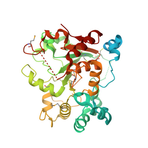
    6APJ, 6APL

  • PubMed Abstract: 

    Vertebrate glycoproteins and glycolipids are synthesized in complex biosynthetic pathways localized predominantly within membrane compartments of the secretory pathway. The enzymes that catalyze these reactions are exquisitely specific, yet few have been extensively characterized because of challenges associated with their recombinant expression as functional products. We used a modular approach to create an expression vector library encoding all known human glycosyltransferases, glycoside hydrolases, and sulfotransferases, as well as other glycan-modifying enzymes. We then expressed the enzymes as secreted catalytic domain fusion proteins in mammalian and insect cell hosts, purified and characterized a subset of the enzymes, and determined the structure of one enzyme, the sialyltransferase ST6GalNAcII. Many enzymes were produced at high yields and at similar levels in both hosts, but individual protein expression levels varied widely. This expression vector library will be a transformative resource for recombinant enzyme production, broadly enabling structure-function studies and expanding applications of these enzymes in glycochemistry and glycobiology.


  • Organizational Affiliation
    • Complex Carbohydrate Research Center, University of Georgia, Athens, Georgia, USA.

Macromolecule Content 

  • Total Structure Weight: 257.78 kDa 
  • Atom Count: 14,650 
  • Modeled Residue Count: 1,772 
  • Deposited Residue Count: 2,244 
  • Unique protein chains: 1

Macromolecules

Find similar proteins by:|  3D Structure
Entity ID: 1
MoleculeChains  Sequence LengthOrganismDetailsImage
Alpha-N-acetylgalactosaminide alpha-2,6-sialyltransferase 2
A, B, C, D, E
A, B, C, D, E, F
374Homo sapiensMutation(s): 0 
Gene Names: ST6GALNAC2SIAT7BSIATL1STHM
EC: 2.4.99 (PDB Primary Data), 2.4.3.3 (UniProt)
UniProt & NIH Common Fund Data Resources
Find proteins for Q9UJ37 (Homo sapiens)
Explore Q9UJ37 
Go to UniProtKB:  Q9UJ37
PHAROS:  Q9UJ37
GTEx:  ENSG00000070731 
Entity Groups
Sequence Clusters30% Identity50% Identity70% Identity90% Identity95% Identity100% Identity
UniProt GroupQ9UJ37
Glycosylation
Glycosylation Sites: 2Go to GlyGen: Q9UJ37-1
Sequence Annotations
Expand
Reference Sequence

Small Molecules

Ligands 2 Unique
IDChains Name / Formula / InChI Key2D Diagram3D Interactions
C5P

Query on C5P


Download:Ideal Coordinates CCD File
G [auth A]
J [auth B]
M [auth C]
P [auth D]
T [auth E]
G [auth A],
J [auth B],
M [auth C],
P [auth D],
T [auth E],
U [auth F]
CYTIDINE-5'-MONOPHOSPHATE
C9 H14 N3 O8 P
IERHLVCPSMICTF-XVFCMESISA-N
NAG

Query on NAG


Download:Ideal Coordinates CCD File
H [auth A]
I [auth A]
K [auth B]
L [auth B]
N [auth C]
H [auth A],
I [auth A],
K [auth B],
L [auth B],
N [auth C],
O [auth C],
Q [auth D],
R [auth D],
S [auth D],
V [auth F]
2-acetamido-2-deoxy-beta-D-glucopyranose
C8 H15 N O6
OVRNDRQMDRJTHS-FMDGEEDCSA-N
Modified Residues  1 Unique
IDChains TypeFormula2D DiagramParent
MSE
Query on MSE
A, B, C, D, E
A, B, C, D, E, F
L-PEPTIDE LINKINGC5 H11 N O2 SeMET

Experimental Data & Validation

Experimental Data

  • Method: X-RAY DIFFRACTION
  • Resolution: 2.35 Å
  • R-Value Free:  0.236 (Depositor), 0.238 (DCC) 
  • R-Value Work:  0.196 (Depositor), 0.199 (DCC) 
  • R-Value Observed: 0.200 (Depositor) 
Space Group: P 1
Unit Cell:
Length ( Å )Angle ( ˚ )
a = 71.168α = 103.69
b = 71.125β = 97.26
c = 138.608γ = 103.03
Software Package:
Software NamePurpose
PHENIXrefinement
HKL-2000data reduction
HKL-2000data scaling
MOLREPphasing

Structure Validation

View Full Validation Report


Ligand Structure Quality Assessment 


Entry History 

Deposition Data

Revision History  (Full details and data files)

  • Version 1.0: 2017-12-20
    Type: Initial release
  • Version 1.1: 2018-01-03
    Changes: Database references
  • Version 1.2: 2018-01-24
    Changes: Database references
  • Version 1.3: 2020-07-29
    Type: Remediation
    Reason: Carbohydrate remediation
    Changes: Data collection, Derived calculations, Structure summary
  • Version 1.4: 2024-04-03
    Changes: Data collection, Database references, Refinement description, Structure summary
  • Version 1.5: 2024-11-20
    Changes: Structure summary