


Crystal Structure of Histidinol Dehydrogenase from Elizabethkingia anophelis
Dranow, D.M., Mayclin, S.J., Lorimer, D.D., Edwards, T.E.To be published.
Experimental Data Snapshot
Starting Model: experimental
View more details
Entity ID: 1 | |||||
|---|---|---|---|---|---|
| Molecule | Chains | Sequence Length | Organism | Details | Image |
| Histidinol dehydrogenase | 447 | Elizabethkingia anophelis NUHP1 | Mutation(s): 0 Gene Names: hisD, BD94_1332 EC: 1.1.1.23 | 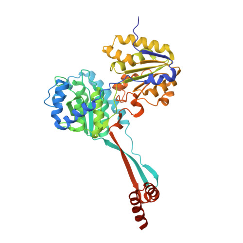 | |
UniProt | |||||
Find proteins for A0A077ECF2 (Elizabethkingia anophelis NUHP1) Explore A0A077ECF2 Go to UniProtKB: A0A077ECF2 | |||||
Entity Groups | |||||
| Sequence Clusters | 30% Identity50% Identity70% Identity90% Identity95% Identity100% Identity | ||||
| UniProt Group | A0A077ECF2 | ||||
Sequence AnnotationsExpand | |||||
Reference Sequence | |||||
| Ligands 4 Unique | |||||
|---|---|---|---|---|---|
| ID | Chains | Name / Formula / InChI Key | 2D Diagram | 3D Interactions | |
| HIS Download:Ideal Coordinates CCD File | U [auth A] | HISTIDINE C6 H10 N3 O2 HNDVDQJCIGZPNO-YFKPBYRVSA-O |  | ||
| IOD Download:Ideal Coordinates CCD File | H [auth A] I [auth A] J [auth A] K [auth A] L [auth A] | IODIDE ION I XMBWDFGMSWQBCA-UHFFFAOYSA-M |  | ||
| ZN Download:Ideal Coordinates CCD File | B [auth A] | ZINC ION Zn PTFCDOFLOPIGGS-UHFFFAOYSA-N |  | ||
| EDO Download:Ideal Coordinates CCD File | C [auth A], D [auth A], E [auth A], F [auth A], G [auth A] | 1,2-ETHANEDIOL C2 H6 O2 LYCAIKOWRPUZTN-UHFFFAOYSA-N |  | ||
| Length ( Å ) | Angle ( ˚ ) |
|---|---|
| a = 82.52 | α = 90 |
| b = 170.08 | β = 90 |
| c = 65.9 | γ = 90 |
| Software Name | Purpose |
|---|---|
| XSCALE | data scaling |
| PHENIX | refinement |
| PDB_EXTRACT | data extraction |
| XDS | data reduction |
| MoRDa | phasing |


