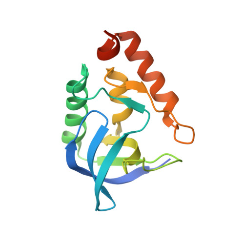
Dielectric Properties of a Protein Probed by Reversal of a Buried Ion Pair.
Robinson, A.C., Schlessman, J.L., Garcia-Moreno E, B.(2018) J Phys Chem B 122: 2516-2524
- PubMed: 29466010
- DOI: https://doi.org/10.1021/acs.jpcb.7b12121
- Primary Citation of Related Structures:
6AMF, 6B8R - PubMed Abstract:
Thirty years ago, Hwang and Warshel suggested that a microenvironment preorganized to stabilize an ion pair would be incapable of reorganizing to stabilize the reverse ion pair. The implications were that (1) proteins have a limited capacity to reorganize, even under the influence of strong interactions, such as those present when ionizable groups are buried in the hydrophobic interior of a protein, and (2) the inability of proteins to tolerate the reversal of buried ion pairs demonstrates the limitations inherent to continuum electrostatic models of proteins. Previously we showed that when buried individually in the interior of staphylococcal nuclease, Glu23 and Lys36 have p K a values near pH 7, but when buried simultaneously, they establish a strong interaction of ∼5 kcal/mol and have p K a values shifted toward more normal values. Here, using equilibrium thermodynamic measurements, crystal structures, and NMR spectroscopy experiments, we show that although the reversed, individual substitutions-Lys23 and Glu36-also have p K a values near 7, when buried together, they neither establish a strong interaction nor promote reorganization of their microenvironment. These experiments both confirm Warshel's original hypothesis and expand it by showing that it applies to reorganization, as demonstrated by our artificial ion pairs, as well as to preorganization as is commonly argued for motifs that stabilize naturally occurring ion pairs in polar microenvironments. These data constitute a challenging benchmark useful to test the ability of structure-based algorithms to reproduce the compensation between self-energy, Coulomb and polar interactions in hydrophobic environments of proteins.
- Department of Biophysics , Johns Hopkins University , Baltimore , Maryland 21218 , United States.
Organizational Affiliation:


