6AL4

CRYSTAL STRUCTURE OF ANTI-CD19 ANTIBODY B43 FAB


Experimental Data Snapshot

  • Method: X-RAY DIFFRACTION
  • Resolution: 2.45 Å
  • R-Value Free: 0.236 
  • R-Value Work: 0.206 
  • R-Value Observed: 0.206 

wwPDB Validation   3D Report Full Report


This is version 2.0 of the entry. See complete history


Literature

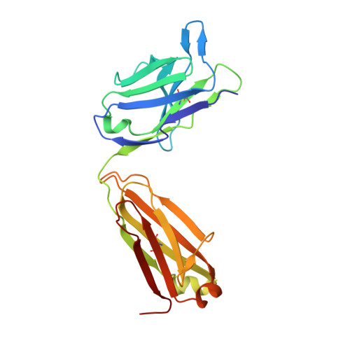
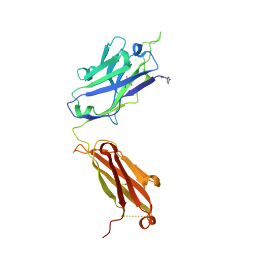
Crystal structure of B-cell co-receptor CD19 in complex with antibody B43 reveals an unexpected fold.

Teplyakov, A.Obmolova, G.Luo, J.Gilliland, G.L.

(2018) Proteins 86: 495-500

  • DOI: https://doi.org/10.1002/prot.25485
  • Primary Citation of Related Structures:  
    6AL4

  • PubMed Abstract: 

    CD19 is a transmembrane protein expressed on malignant B cells, but not in other lineages or other tissues, which makes it an attractive target for monoclonal antibody-mediated immunotherapy. Anti-CD19 antibody B43 was utilized in a bispecific T-cell engager (BiTE) blinatumomab that demonstrated potency for the treatment of relapsed acute lymphoblastic leukemia. To gain insight into the mechanism of action of the antibody, the crystal structure of B43 Fab was determined in complex with CD19 and in the unbound form. The structure revealed the binding epitope, explained the lack of cross-reactivity toward non-human species, and suggested the key-and-lock mechanism of antigen recognition. Most unexpectedly, the structure revealed a unique molecular topology of CD19. Rather than a tandem of c-type immunoglobulin folds predicted from the amino acid sequence, the extracellular domain of CD19 exhibits an elongated β-sandwich formed by two immunoglobulin folds by swapping their C-terminal halves. This is the first structure of CD19, which has no sequence homologs.


  • Organizational Affiliation

    Janssen Research and Development, LLC, 1400 McKean Road, Spring House, Pennsylvania, 19477.


Macromolecules
Find similar proteins by:  (by identity cutoff)  |  3D Structure
Entity ID: 1
MoleculeChains Sequence LengthOrganismDetailsImage
B43 LIGHT CHAIN
A, C, E
218Homo sapiensMutation(s): 0 
UniProt
Find proteins for Q6P5S8 (Homo sapiens)
Explore Q6P5S8 
Go to UniProtKB:  Q6P5S8
Entity Groups  
Sequence Clusters30% Identity50% Identity70% Identity90% Identity95% Identity100% Identity
UniProt GroupQ6P5S8
Sequence Annotations
Expand
  • Reference Sequence
Find similar proteins by:  (by identity cutoff)  |  3D Structure
Entity ID: 2
MoleculeChains Sequence LengthOrganismDetailsImage
B43 HEAVY CHAIN
B, D, F
233Homo sapiensMutation(s): 0 
UniProt
Find proteins for Q6GMX6 (Homo sapiens)
Explore Q6GMX6 
Go to UniProtKB:  Q6GMX6
Entity Groups  
Sequence Clusters30% Identity50% Identity70% Identity90% Identity95% Identity100% Identity
UniProt GroupQ6GMX6
Sequence Annotations
Expand
  • Reference Sequence
Small Molecules
Modified Residues  1 Unique
IDChains TypeFormula2D DiagramParent
PCA
Query on PCA
B, D, F
L-PEPTIDE LINKINGC5 H7 N O3GLN
Experimental Data & Validation

Experimental Data

  • Method: X-RAY DIFFRACTION
  • Resolution: 2.45 Å
  • R-Value Free: 0.236 
  • R-Value Work: 0.206 
  • R-Value Observed: 0.206 
  • Space Group: P 21 21 21
Unit Cell:
Length ( Å )Angle ( ˚ )
a = 73.1α = 90
b = 127.36β = 90
c = 219.39γ = 90
Software Package:
Software NamePurpose
HKL-2000data reduction
HKL-2000data scaling
REFMACrefinement
PHASERphasing

Structure Validation

View Full Validation Report


Entry History 

Deposition Data

Revision History  (Full details and data files)

  • Version 1.0: 2018-03-28
    Type: Initial release
  • Version 1.1: 2018-05-02
    Changes: Data collection, Database references
  • Version 2.0: 2023-10-04
    Changes: Data collection, Database references, Polymer sequence, Refinement description