6AII | pdb_00006aii

Catalytic domain of PdAgaC


Experimental Data Snapshot

  • Method: X-RAY DIFFRACTION
  • Resolution: 1.63 Å
  • R-Value Free: 
    0.198 (Depositor), 0.202 (DCC) 
  • R-Value Work: 
    0.174 (Depositor), 0.186 (DCC) 
  • R-Value Observed: 
    0.175 (Depositor) 

Starting Model: experimental
View more details

wwPDB Validation 3D Report Full Report

Validation slider image for 6AII

This is version 1.2 of the entry. See complete history

Literature

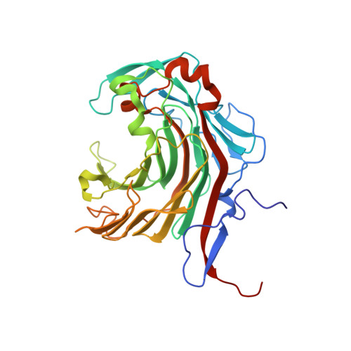
Crystal structure of a neoagarobiose-producing GH16 family beta-agarase from Persicobacter sp. CCB-QB2.

Teh, A.H.Fazli, N.H.Furusawa, G.

(2020) Appl Microbiol Biotechnol 104: 633-641

  • DOI: https://doi.org/10.1007/s00253-019-10237-y
  • Primary Citation Related Structures: 
    6AII

  • PubMed Abstract: 

    PdAgaC from the marine bacterium Persicobacter sp. CCB-QB2 is a β-agarase belonging to the glycoside hydrolase family 16 (GH16). It is one of only a handful of endo-acting GH16 β-agarases able to degrade agar completely to produce neoagarobiose (NA2). The crystal structure of PdAgaC's catalytic domain, which has one of the highest V max value at 2.9 × 10 3 U/mg, was determined in order to understand its unique mechanism. The catalytic domain is made up of a typical β-jelly roll fold with two additional insertions, and a well-conserved but wider substrate-binding cleft with some minor changes. Among the unique differences, two unconserved residues, Asn226 and Arg286, may potentially contribute additional hydrogen bonds to subsites -1 and +2, respectively, while a third, His185 from one of the additional insertions, may further contribute another bond to subsite +2. These additional hydrogen bonds may probably have enhanced PdAgaC's affinity for short agaro-oligosaccharides such as neoagarotetraose (NA4), rendering it capable of binding NA4 strongly enough for rapid degradation into NA2.


  • Organizational Affiliation
    • Centre for Chemical Biology, Universiti Sains Malaysia, 10 Persiaran Bukit Jambul, 11900 Bayan Lepas, Penang, Malaysia. aikhong@usm.my.

Macromolecule Content 

  • Total Structure Weight: 38.48 kDa 
  • Atom Count: 3,045 
  • Modeled Residue Count: 330 
  • Deposited Residue Count: 334 
  • Unique protein chains: 1

Macromolecules

Find similar proteins by:|  3D Structure
Entity ID: 1
MoleculeChains  Sequence LengthOrganismDetailsImage
Beta-agarase334PersicobacterMutation(s): 0 
UniProt
Find proteins for A0A5S8WF47 (Persicobacter)
Explore A0A5S8WF47 
Go to UniProtKB:  A0A5S8WF47
Entity Groups
Sequence Clusters30% Identity50% Identity70% Identity90% Identity95% Identity100% Identity
UniProt GroupA0A5S8WF47
Sequence Annotations
Expand
Reference Sequence

Small Molecules

Ligands 1 Unique
IDChains Name / Formula / InChI Key2D Diagram3D Interactions
CA

Query on CA


Download:Ideal Coordinates CCD File
B [auth A]CALCIUM ION
Ca
BHPQYMZQTOCNFJ-UHFFFAOYSA-N

Experimental Data & Validation

Experimental Data

  • Method: X-RAY DIFFRACTION
  • Resolution: 1.63 Å
  • R-Value Free:  0.198 (Depositor), 0.202 (DCC) 
  • R-Value Work:  0.174 (Depositor), 0.186 (DCC) 
  • R-Value Observed: 0.175 (Depositor) 
Space Group: P 41 21 2
Unit Cell:
Length ( Å )Angle ( ˚ )
a = 76.779α = 90
b = 76.779β = 90
c = 166.05γ = 90
Software Package:
Software NamePurpose
REFMACrefinement
d*TREKdata reduction
d*TREKdata scaling
MOLREPphasing

Structure Validation

View Full Validation Report


Entry History 

& Funding Information

Deposition Data


Funding OrganizationLocationGrant Number
Malaysia--

Revision History  (Full details and data files)

  • Version 1.0: 2019-08-28
    Type: Initial release
  • Version 1.1: 2020-04-08
    Changes: Database references
  • Version 1.2: 2023-11-22
    Changes: Data collection, Database references, Refinement description