


Crystal structure of enoyl-ACP reductase from Acinetobacter baumannii in complex with NAD and Triclosan
Rani, S.T., Nataraj, V., Laxminarasimhan, A., Thomas, A., Krishnamurthy, N.To be published.
Experimental Data Snapshot
Starting Model: experimental
View more details
Entity ID: 1 | |||||
|---|---|---|---|---|---|
| Molecule | Chains | Sequence Length | Organism | Details | Image |
| Enoyl-[acyl-carrier-protein] reductase [NADH] | 267 | Acinetobacter baumannii ATCC 19606 = CIP 70.34 = JCM 6841 | Mutation(s): 0 Gene Names: NC_009085.1 EC: 1.3.1.9 | 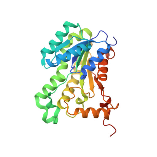 | |
UniProt | |||||
Entity Groups | |||||
| Sequence Clusters | 30% Identity50% Identity70% Identity90% Identity95% Identity100% Identity | ||||
| UniProt Group | D0CAD5 | ||||
Sequence AnnotationsExpand | |||||
Reference Sequence | |||||
| Ligands 2 Unique | |||||
|---|---|---|---|---|---|
| ID | Chains | Name / Formula / InChI Key | 2D Diagram | 3D Interactions | |
| NAD Download:Ideal Coordinates CCD File | B [auth A] | NICOTINAMIDE-ADENINE-DINUCLEOTIDE C21 H27 N7 O14 P2 BAWFJGJZGIEFAR-NNYOXOHSSA-N |  | ||
| TCL Download:Ideal Coordinates CCD File | C [auth A] | TRICLOSAN C12 H7 Cl3 O2 XEFQLINVKFYRCS-UHFFFAOYSA-N |  | ||
| Length ( Å ) | Angle ( ˚ ) |
|---|---|
| a = 77.212 | α = 90 |
| b = 77.245 | β = 90 |
| c = 85.207 | γ = 90 |
| Software Name | Purpose |
|---|---|
| REFMAC | refinement |
| XDS | data reduction |
| SCALEPACK | data scaling |
| MOLREP | phasing |


