6AAV | pdb_00006aav

Crystal structure of alpha-glucosyl transfer enzyme, XgtA at 1.72 angstrom resolution


Experimental Data Snapshot

  • Method: X-RAY DIFFRACTION
  • Resolution: 1.72 Å
  • R-Value Free: 
    0.254 (Depositor), 0.257 (DCC) 
  • R-Value Work: 
    0.218 (Depositor), 0.220 (DCC) 
  • R-Value Observed: 
    0.220 (Depositor) 

Starting Model: experimental
View more details

wwPDB Validation 3D Report Full Report

Validation slider image for 6AAV

This is version 1.3 of the entry. See complete history

Literature

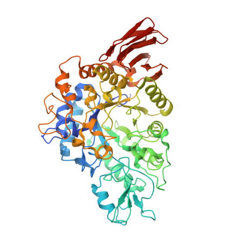
Crystal structure of alpha-glucosyl transfer enzyme XgtA from Xanthomonas campestris WU-9701.

Watanabe, R.Arimura, Y.Ishii, Y.Kirimura, K.

(2020) Biochem Biophys Res Commun 526: 580-585

  • DOI: https://doi.org/10.1016/j.bbrc.2020.03.109
  • Primary Citation Related Structures: 
    6AAV

  • PubMed Abstract: 

    The α-glucosyl transfer enzyme XgtA is a novel type α-Glucosidase (EC 3.2.1.20) produced by Xanthomonas campestris WU-9701. One of the unique properties of XgtA is that it shows extremely high α-glucosylation activity toward alcoholic and phenolic -OH groups in compounds using maltose as an α-glucosyl donor and allows for the synthesis of various useful α-glucosides with high yields. XgtA shows no hydrolytic activity toward sucrose and no α-glucosylation activity toward saccharides to produce oligosaccharides. In this report, the crystal structure of XgtA was solved at 1.72 Å resolution. The crystal belonged to space group P22 1 2 1 , with unit-cell parameters a = 73.07, b = 83.48, and c = 180.79 Å. The β→α loop 4 of XgtA, which is proximal to the catalytic center, formed a unique structure that is not observed in XgtA homologs. Furthermore, XgtA was found to contain unique amino acid residues around its catalytic center. The unique structure of XgtA provides an insight into the mechanism for the regulation of substrate specificity in this enzyme.


  • Organizational Affiliation
    • Department of Applied Chemistry, Faculty of Science and Engineering, Waseda University, 3-4-1 Ohkubo, Shinjuku-ku, Tokyo, 169-8555, Japan.

Macromolecule Content 

  • Total Structure Weight: 121.27 kDa 
  • Atom Count: 9,068 
  • Modeled Residue Count: 1,071 
  • Deposited Residue Count: 1,088 
  • Unique protein chains: 1

Macromolecules

Find similar proteins by:|  3D Structure
Entity ID: 1
MoleculeChains  Sequence LengthOrganismDetailsImage
Alpha-glucosyltransferase
A, B
544Xanthomonas campestrisMutation(s): 0 
Gene Names: xgtA
UniProt
Find proteins for Q76LB0 (Xanthomonas campestris)
Explore Q76LB0 
Go to UniProtKB:  Q76LB0
Entity Groups
Sequence Clusters30% Identity50% Identity70% Identity90% Identity95% Identity100% Identity
UniProt GroupQ76LB0
Sequence Annotations
Expand
Reference Sequence

Experimental Data & Validation

Experimental Data

  • Method: X-RAY DIFFRACTION
  • Resolution: 1.72 Å
  • R-Value Free:  0.254 (Depositor), 0.257 (DCC) 
  • R-Value Work:  0.218 (Depositor), 0.220 (DCC) 
  • R-Value Observed: 0.220 (Depositor) 
Space Group: P 2 21 21
Unit Cell:
Length ( Å )Angle ( ˚ )
a = 73.071α = 90
b = 83.478β = 90
c = 180.785γ = 90
Software Package:
Software NamePurpose
XDSdata reduction
Aimlessdata scaling
PHASERphasing
PHENIXrefinement
PDB_EXTRACTdata extraction

Structure Validation

View Full Validation Report


Entry History 

Deposition Data

Revision History  (Full details and data files)

  • Version 1.0: 2019-07-24
    Type: Initial release
  • Version 1.1: 2020-05-13
    Changes: Database references
  • Version 1.2: 2020-05-20
    Changes: Database references
  • Version 1.3: 2023-11-22
    Changes: Data collection, Database references, Refinement description